
Buspar dosages: 10 mg, 5 mg
Buspar packs: 90 pills, 120 pills, 180 pills, 270 pills, 360 pills

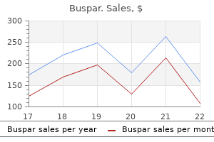
Purchase 10 mg buspar visa
The first-formed dentine anxiety symptoms gi 10 mg buspar purchase, the mantle dentine anxiety symptoms 97 order buspar 10 mg without a prescription, might include some parts derived from subodontoblastic cells. The sample of innervation of the dental pulp is established in the course of the cap stage of tooth development. Mineralization of dentine happens initially by the budding off of matrix vesicles from the odontoblast cell. Like enamel, maturation of dentine commences when the complete thickness of the dentine has been formed. To attain the mineralizing front, hydroxyapatite crystals cross through the odontoblast cells. A pool of chondroitin sulphate proteoglycans is secreted at the mineralization front and guides mineral deposition. It stains in another way compared with the natural element of mineralized dentine 181 Thirteen: Dental tissues. Some developmental features may be seen in the dentine of a completely formed tooth considered under polarized mild. Identify the rounded/arcade outlines (A) and the dark line (B) and explain their significance. This is a scanning electron micrograph of the surface of dentine after it has been handled with hypochlorite. The outline of the mineralization entrance in dentine is usually seen as a combination of each types of mineralization. There is commonly much attrition and the condition is generalized across all enamel within the dentition. It is thus likely to be thicker in younger tooth where the pace of formation is fast, and thinner within the enamel of older sufferers the place the pace of formation is slower. Dentine phosphophoryn (thought to be important in mineralization) is transported alongside the odontoblast course of, bypassing the predentine, and is deposited at the mineralizing entrance. Calcium and other elements pass separately through (or between) the odontoblasts in their ionic state. As calcium is probably poisonous to cells, the odontoblast cell possesses numerous mechanisms to transport this factor via the cell without inflicting hurt. They are thought to direct mineral deposition and are secreted by odontoblasts on the mineralization entrance solely. The presence of enamel-related proteins during regular root dentine formation underlies the explanation for making use of related proteins to root surfaces in makes an attempt to improve the success price of periodontal regeneration. This distinction in staining is the outcomes of biochemical modifications occurring on the mineralizing entrance, involving the addition of new biochemical molecules and the degradation of others. They may be considerably elevated in pathological conditions affecting mineralization, such as hypophosphataemia. The matrix vesicles have many molecules favouring the initiation of mineralization within them, similar to alkaline phosphatase and calcium-binding proteins. Due to the radial orientation of the enamel crystallites in calcospherites, the extent of this pattern of dentine mineralization can be distinguished from the linear kind by viewing a ground section in polarized mild. Unlike the adult condition, growing enamel matrix is retained in demineralized sections due to its higher natural content material. The two tissues stain in a different way due to their different compositions, dentine being primarily collagenous, while enamel matrix consists of enamel proteins (mainly amelogenins and non-amelogenins). Polarizing microscopy can reveal differences within the orientation of collagen and hydroxyapatite crystals. The rounded/arcade outlines (A) indicate the radial orientation of hydroxyapatite crystals found in 183 Thirteen: Dental tissues. The collagen fibrils in mantle dentine are oriented perpendicular to the enamel� dentine junction, whereas those in circumpulpal dentine are oriented parallel to the enamel�dentine junction. The Tomes strategy of the ameloblast stays at the mineralizing enamel entrance and is responsible for producing the prismatic construction. The natural matrix of dentine is principally collagen, which is little changed during formation. However, the organic matrix of enamel is comprised of unique enamel proteins that undergo degradation and are largely lost in the course of the maturation of enamel. In this fashion, enamel reaches a higher diploma of mineralization and its crystals are bigger. Enamel, being restricted to the crown, has completed its formation nicely earlier than the tooth erupts, though some post-eruptive maturation occurs within the surface layers on account of ionic change with saliva. Enamel, being non-vital, reveals little response, although small initial carious lesions could remineralize beneath suitable conditions. The utility of hypochlorite removes the natural material (predentine) covering the mineralizing dentine. The first-formed dentine (mantle dentine) mineralizes inside matrix vesicles budded off from the odontoblast. This is a section of the crown from a affected person who, due to illness, has obtained 5 separate injections of tetracycline. This drug is taken up after which localized at the mineralizing entrance of dentine (arrows) on every event, which may be visualized using fluorescence microscopy. Many other markers can be integrated into the mineralizing front, similar to alizarin red, as well as autographic techniques (using tritiated glycine or proline that turns into integrated into developing collagen). Outline essay solutions Question 1 the main options to be talked about are as follows: � There are reciprocal interactions between enamel- and dentine-forming cells throughout growth. Question 2 the fundamental unit of dentine seen in a floor section is the dentinal tubule. As odontoblasts on the periphery of the pulp migrate pulpwards (centripetally), they path behind them the odontoblast processes. From their distal surfaces, the odontoblasts secrete the natural matrix of dentine across the processes. This matrix consists of collagen fibrils and the related ground substance, and constitutes the predentine layer. In the initial mantle layer, the collagen fibrils are oriented perpendicular to the enamel� dentine junction. This contrasts with the relaxation of the circumpulpal dentine where the collagen is oriented parallel to 184 Thirteen: Dental tissues. Thus, the mantle layer can be distinguished if a ground section of the crown is considered in polarized gentle. When this predentine matures, mineralization happens, leading to tubule formation. The odontoblasts within the crown migrate centrally (centripetally) and gradually occupy a smaller surface area; in doing so, they hint out a sinusoidal path, giving the primary curvatures of the tubules.
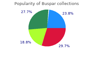
Buspar 10 mg buy on line
A plain skull x-ray has been ordered usually by physicians apart from cranium base surgeons or junior colleagues anxiety symptoms 8 year old boy buspar 10 mg generic line. Any skull base lesion that has a large demanding blood supply could cause asymmetry of the foramina by way of which principle vessels traverse anxiety symptoms videos buspar 10 mg buy generic, such because the foramen spinosum. Failure to identify such asymmetry within the presence of tinnitus and to prepare additional radiological investigations has been thought of substandard care. These sufferers are sometimes in early middle age and as such merit diligent investigation of their tinnitus, notably if they describe it as being loud and located to one particular a half of the top. There is little question that angiography is the gold commonplace, however in contemplating its use the risks need to be thought of, defined to the patient and knowledgeable consent given. These happen in 3�14 p.c of revealed sequence and may be regularly associated with meningitis. Postoperative intracranial bleeding is probably the most feared and acute complication and it behoves the surgeon to have a technique to manage the state of affairs. Neurosurgical centres have surgical workers on site to decompress the area instantly by opening the wound. Temporal paragangliomas When small, the one symptom of these lesions is tinnitus. However, often decrease cranial nerve palsy can happen with out apparent middle ear disease. Describe the muscle attachments for the mandible and indicate how knowledge of these attachments could aid our understanding of the displacement of bony fragments following fractures of the physique of the mandible. The opening of the parotid duct might present as a papilla or as a simple opening into the cheek. The soft palate is raised during swallowing by the mixed actions of the levator and tensor veli palatini muscle tissue. Competent lips produce an anterior oral seal and ensure the right inclination of the incisors because the competent decrease lip pushes in opposition to each the lower and the upper incisors. The infra-orbital foramen does transmit the infra-orbital nerve (and related blood vessels) however this nerve is a department of the maxillary division of the trigeminal. The superior genial tubercles give rise to the genioglossus muscular tissues and the inferior tubercles give rise to the geniohyoid muscle tissue. The anterior bellies of the digastric muscles are attached to the digastric fossae under the genial tubercles and at the inferior border of the mandible. The mylohyoid muscles (attached collectively at the midline by a raphe) kind the diaphragm for the ground of the mouth. The palatine raphe within the centre of the onerous palate is firmly certain to the underlying bone (forming a mucoperiosteum). It is alleged that the mental foramen normally lies beneath the roots of a premolar tooth. The opening of the maxillary air sinus (ostium) lies high up in the course of the roof of the sinus (an unfavourable location for drainage of the mucus into the lateral wall of the nose, hiatus semilunaris of the center meatus). The pterygomandibular raphe extends from the pterygoid hamulus to the retromolar fossa behind the mandibular third molar tooth. The two mylohyoid muscle tissue collectively type the diaphragm for the mouth and delineate the floor of the mouth from the suprahyoid area of the neck (although both regions communicate on the posterior edges of the diaphragm). Tori mandibulares are bony exostoses extending from the mandibular alveolus into the area of the ground of the mouth. The mucosa over the hard palate has a keratinized (masticatory) stratified squamous epithelium. The nasopalatine nerves exit on the incisive fossa and are thus located under the incisive papilla and behind the maxillary central incisor enamel. The larger palatine nerves run in a submucosa, every along a lateral channel between the maxillary alveolar and the maxillary palatine processes. The mucoperiosteum is an effective barrier to the spread of infection from the maxillary teeth into the onerous palate. At G, the lingula, is attached the sphenomandibular ligament (an accessory ligament of the temporomandibular joint). At A, the genial spines, are attached the genioglossus muscle tissue (superior spines) and geniohyoid muscular tissues (inferior spines). At B, the mylohyoid ridge, is connected the mylohyoid muscle which contributes to the diaphragm for the ground of the mouth. At C, the internal side of the angle of the mandible, is connected the medial pterygoid muscle. At I, the digastric fossa, is attached the anterior belly of the digastric muscle. The lingual department of the mandibular nerve runs on the lingual alveolar plate of the permanent mandibular third molar tooth and must subsequently be protected during surgical extraction of this tooth. If the nerve is broken, there will be: (i) lack of basic sensation to the tongue (ventral and dorsal surfaces), ground of mouth and lingual gingivae; (ii) lack of particular sensation (taste) to the anterior two-thirds of the tongue; and (iii) loss of secretomotor provide to the submandibular and sublingual salivary glands. The losses associated with (ii) and (iii) outcome from damage to the fibres from the chorda tympani department of the facial nerve (nervus intermedius) which passes with the lingual nerve. We know the radiograph is of an anatomical specimen due to the absence of the vertebral column. F is the ridge produced by the pterygomandibular raphe, which passes from the pterygoid hamulus to the posterior end of the mylohyoid line. Its scientific significance is as a landmark for an inferior alveolar nerve block (see web page 69). Four of the muscle tissue of the palate (palatoglossus, palatopharyngeus, musculus uvulae and levator veli palatini) derive their nerve supply from the pharyngeal plexus, while the remaining muscle (tensor veli palatini) is provided by a department of the mandibular nerve. The sensory supply to the soft palate is by way of the greater and lesser palatine nerves. Minor salivary glands in the soft palate derive their secretomotor supply from the higher petrosal nerve by way of the pterygopalatine ganglion. At D, the incisive fossa, emerge the nasopalatine nerves by way of the pterygopalatine ganglion; these are branches of the maxillary nerve. At F, the higher palatine foramen, emerge the larger palatine nerve, also from the maxillary nerve through the pterygopalatine ganglion, and the greater palatine artery, a department from the third part of the maxillary artery. At G, the lesser palatine foramen, emerge the lesser palatine nerves, again from the maxillary nerve through the pterygopalatine ganglion, accompanied by the lesser palatine artery, a branch from the third a half of the maxillary artery. Developmentally, the mouth is a area the place each ectoderm and endoderm contribute. Hormonal changes at puberty stimulate the formation and secretion of sebaceous glands. Outline essay solutions Question 1 Writing essays requires a logical ordering of data and, wherever possible, some proof of study and important considering. A concluding paragraph ought to be supplied that summarizes the essential/important variations. Question 2 the introduction to this essay ought to be basic and brief, highlighting essential details about the mandible, the temporomandibular joint and the process of mastication (a attribute of mammals). The essay must be structured in order that the muscle tissue are categorised in accordance with the final subdivision of mandible right into a physique and the rami (see Table 1.
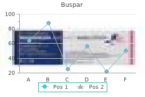
5 mg buspar generic otc
This area is attribute of the individual and might be maintained by resting muscle length that controls mandibular posture anxiety frequent urination purchase 5 mg buspar otc. The curve of Spee describes the curvatures of the dental arches in the sagittal (anteroposterior) aircraft anxiety management 5 mg buspar discount with amex. The curves of Wilson describe the curvatures of the posterior teeth within the coronal (transverse) plane. The curves of Monson (often erroneously used to describe the curves of Wilson) combine each curves as if the teeth rested on a segment of a sphere of about 10 cm. The maxillary arch is barely bigger and broader than the mandibular arch and due to this fact usually overlaps the mandibular arch by a couple of millimetres. Overbite refers to the way the maxillary incisors and canine vertically overlap the labial surfaces of the mandibular incisors and canine. Crossbites are incessantly associated to discrepancies in the widths of the dental bases and should involve the displacement of the mandible to one side to obtain maximal intercuspation. A Carabelli trait (tubercle and even an extra cusp) is most commonly found on the everlasting maxillary first molar tooth (60% of teeth). A canine fossa is found on the mesial floor of the everlasting maxillary first premolar tooth. Also on this floor is a canine groove (a continuation of the occlusal fissure across the mesial marginal ridge). The notched incisal margin, containing three mammelons, is normal within the just lately erupted tooth however is misplaced with attrition. This may be clinically important throughout endodontic treatment or tooth extraction. The wide-open pulp and the graceful, thin lining of dentine indicate that the basis is incomplete and nonetheless forming. Structure A is a supernumerary tooth which has erupted into the palate behind the everlasting incisors. It might remain unerupted and be discovered on a radiograph when the affected person presents with delayed eruption of the permanent maxillary incisor(s). As evident from the fully erupted first everlasting molars, the affected person is about 9 years of age. Posterior and center superior alveolar nerves, buccal nerve, higher palatine nerve. There ought to follow a description of root morphology for each of the permanent incisors and canines (maxillary and mandibular), with applicable diagrams. The root morphology of the permanent premolars and molars (maxillary and mandibular) should be described, with applicable diagrams. Comparisons of deciduous versus permanent root morphologies can be briefly outlined. General descriptions of the cervical margins (where anatomical crown meets anatomical root) can be offered, again with applicable diagrams. The last paragraph(s) should spotlight some medical concerns, for example: � � � � � � � ageing pulp inflammation dental abscesses root canal remedy must avoid pulps during conservation therapy pulpectomies others. This patient has an anterior open chunk, the mandibular incisors not being overlapped (overbite) by the maxillary incisors. The condition could also be related to an anterior tongue thrust on swallowing, or the patient may be a ordinary thumb sucker. It may be associated with an abnormal and premature occlusal contact on the posterior teeth. It may be associated to underdevelopment of the anterior section of the maxillae. The last paragraph should emphasize the controversies and difficulties outlined in the physique of the essay and will end by discussing whether or not malocclusions are pathological or normal variations. Outline essay answers Question 1 the introductory paragraph ought to provide some basic information regarding the human dentition. There must also be a definition of molars and a short description of their features. A description of the final variations between deciduous and everlasting teeth should observe, then descriptions of specific variations between deciduous and everlasting molars (including numbers and site, chronology of growth, and crown and root morphologies). Mention should be made of the fact that deciduous molars are replaced by permanent premolars and not molars. Question 2 the introductory paragraph should outline roots (anatomical and medical definitions) and mention the tissues comprising the roots (together with a diagram). All the muscular tissues of mastication receive their innervation from the mandibular division of the trigeminal nerve. Closely associated functionally with the muscles of mastication is the digastric muscle. The masseter and temporalis muscles lie on the superficial face, whereas the lateral and medial pterygoid muscles lie deeper inside the infratemporal fossa. Masseter Overview Extra-orally, the muscular tissues of mastication transfer the mandible on the temporomandibular joint while the circumoral muscular tissues of facial expression change the shapes and positions of the lips. In the suprahyoid area, the digastric, mylohyoid and geniohyoid muscular tissues are situated within the flooring of the mouth. Intraorally, the taste bud (the movable part of the palate) is raised and elevated by muscle tissue throughout and after swallowing and the form and place of the tongue is affected by intrinsic and extrinsic musculature (see pages 52�53). Chewing (mastication) and swallowing (deglutition) are necessary functions involving the orofacial musculature. The masseter muscle consists of two overlapping heads: � the superficial head arises from the zygomatic strategy of the maxilla and from the anterior two-thirds of the decrease border of the zygomatic arch. Internally, the muscle has many tendinous septa that significantly increase the realm for muscle attachment and which give a multipennate association, thereby growing its power. The superficial head passes downwards and backwards to insert into the decrease half of the lateral surface of the ramus. The deep head, whose posterior fibres are extra vertically oriented, inserts into the higher half of the lateral floor of the ramus, notably over the coronoid course of. The muscle elevates the mandible and is primarily energetic when grinding powerful meals. Indeed, the muscle exerts appreciable energy when the mandible is near the centric occlusal position. On the premise of its fibre orientation, the posterior fibres of the deep head may have some retrusive functionality for the mandible. Learning aims You should: � have the flexibility to describe the areas, attachments, capabilities and innervations of the muscles influencing mandibular actions and movements of the lips, cheeks and ground of the mouth, and the taste bud (for the musculature of the tongue, see pages 52�53) � perceive the physiological mechanisms underlying the processes (and control) of mastication and swallowing. It takes origin from the floor of the temporal fossa of the lateral floor of the skull and from the overlying temporal fascia, and may thus be considered a bipennate muscle. From this broad origin, the fibres converge towards their insertion on the apex, the anterior and posterior borders, and the medial floor of the coronoid process. Indeed, the insertion extends down the anterior border of the ramus virtually so far as the third molar tooth. The posterior fibres of the muscle cross horizontally forwards while the anterior fibres cross vertically downwards on to the coronoid process.
Order 5 mg buspar with visa
Enamel prisms the fundamental structural unit of enamel is the enamel prism (rod) anxiety symptoms headache buspar 10 mg purchase mastercard, operating from the enamel�dentine junction to the floor anxiety symptoms eyesight 5 mg buspar cheap with mastercard. In a cross-section of human enamel, the prisms may be seen to be keyhole-shaped and alternate, so that the tail of a prism lies between the heads of two prisms in the row beneath. Adjacent prisms are delineated by the prism boundary, an optical function produced by sudden modifications in crystallite orientation at that web site. Although within the outer third of enamel prisms run parallel to each other when viewed in a longitudinal section of the crown, in the internal two-thirds adjacent bands of enamel roughly 50 m broad (and containing groups of about 10�20 prisms) present prisms operating in different instructions as they spiral outwards; some groups of prisms are reduce more Perikymata grooves and ridges Over the entire of the lateral enamel, enamel striae attain the surface in a series of fine grooves operating circumferentially around the crown. These grooves are known as the perikymata grooves and are separated by ridges, the perikymata ridges. In deciduous teeth, enamel striae and perikymata are only ever clearly seen in the cervical enamel of deciduous second molars. Neonatal line Enamel striae are less pronounced or absent from enamel shaped earlier than delivery. A particularly marked stria is formed at birth � this is the neonatal line and reflects the metabolic 143 Twelve: Dental tissues. Enamel formation Overview Being epithelial in origin, enamel formation differs in lots of respects from that related to the opposite mineralized dental tissues. When initially shaped, young enamel is only frivolously mineralized (about 20�30%) and accommodates a excessive proportion of distinctive enamel proteins. However, it subsequently undergoes a process of maturation whereby its very high level of mineral content (96%) is attained and extra enamel proteins and water are eliminated. The extra complicated pattern of improvement of enamel is mirrored in the altering morphology of the ameloblast during growth. Compared with subsurface enamel, surface enamel is tougher, less porous, much less soluble and more radio-opaque. It is richer in some trace elements (especially fluoride) but incorporates less carbonate. The enamel floor presents a variable appearance, exhibiting options similar to aprismatic enamel, perikymata, prism-end markings, cracks, pits and elevations. Learning aims Enamel�dentine junction the boundary between enamel and dentine is identified as the enamel�dentine junction. It is scalloped and this feature is especially evident beneath cusps and incisal edges. Features seen at the enamel�dentine junction embody enamel spindles, enamel tufts and enamel lamellae. You should: � know the completely different levels that occur during enamel formation and be capable of relate the altering construction of the ameloblast with its altering capabilities � respect the composition of the organic matrix and the way this changes during enamel formation � perceive how the structural features noticed within the grownup tissues are associated to the event of the tissue � be capable of evaluating and contrasting enamel and dentine formation. Enamel spindles these are slim, club-shaped structures extending up to 25 m into the enamel; they might characterize odontoblast processes that, through the early levels of enamel growth, insinuate themselves between the ameloblasts. Enamel spindles are mostly seen beneath cusps and, as a outcome of their alignment, are best considered in longitudinal sections of enamel. Enamel formation (amelogenesis) commences at the late bell stage of tooth formation, the earlier modifications having been described in Chapter 10. During the early bell stage, the enamel organ comprises four distinct layers: � � � � External enamel epithelium Stellate reticulum Stratum intermedium Internal enamel epithelium. Enamel tufts these are extra in depth than enamel spindles and are seen in the inside third of the enamel. Resembling tufts of grass, they appear to travel in the same direction as the prisms. The prism boundaries in the tufts are hypomineralized and include extra enamel protein. They recur at roughly 100 m intervals along the enamel�dentine junction and, owing to their alignment, are greatest visualized in transverse sections of enamel. Enamel lamellae these are skinny, sheet-like faults that run through the entire thickness of the enamel. Prior to the formation of dentine and enamel, the shape of the tooth has already been outlined following epithelial/ mesenchymal interactions throughout tooth morphogenesis. The peripheral cells of the dental papilla adjacent to the internal enamel epithelium are undifferentiated, while the inner enamel epithelial cells have assumed a columnar look. During the formation of enamel, the interior enamel epithelial cell undergoes numerous modifications in its morphology, every being related to completely different functions. These could be thought-about for convenience in 5 levels: presecretory, secretory, transitional, maturation and post-maturation. Presecretory stage In the stage leading as much as the secretion of the enamel matrix, the presecretory stage, initial indicators of differentiation happen at the cusp-tip or incisal edge and steadily spread down the perimeters of the crown. There is a build-up of intracellular organelles associated with protein synthesis. Although no extracellular enamel matrix is present at this stage, small quantities of enamel proteins are synthesized and may be involved in epithelial/mesenchymal interactions. Increments of enamel are deposited on one another and enamel formation extends from the cusp-tips down the edges of the tooth. Just earlier than the enamel reaches its last thickness, the Tomes course of disappears and the distal floor of the ameloblast becomes flattened, so that the final 20�100 m at the surface is prismless. The advanced paths traced out by the ameloblasts as they transfer outwards are liable for generating the Hunter�Schreger bands, with groups of prisms in adjoining layers of enamel moving in different instructions. The secretory phase ends once the total thickness of enamel matrix has been laid down. Twelve Secretory stage the secretory stage is characterised by the synthesis and secretion of the enamel matrix and its initial gentle mineralization. At the ultrastructural level, there is a rise in endoplasmic reticulum as nicely as vesicles containing material representing the organic matrix of enamel. The contents of the vesicles (secretory granules) are discharged into the extracellular space on the distal end of the cell towards the floor of the first-formed dentine. Almost as soon as the enamel matrix is launched extracellularly, the preliminary calcium hydroxyapatite crystallites appear inside it as skinny, needle-like crystallites. As the ameloblasts migrate outwards (centrifugally), small processes from the odontoblasts could get caught up between them. When the early enamel starts to mineralize round them, these processes will turn out to be entrapped as enamel spindles. Incremental markings Periodic adjustments within the nature or orientation of the enamel crystallites or enamel matrix or enamel prisms produce short-period or long-period incremental markings. A diurnal rhythm produces a daily cross-striation across each prism roughly 4 m aside, whereas approximately every 7 days (range 6�10), an enamel stria (of Retzius) is produced outlining the mineralizing front and working obliquely to the floor. These striae end on the floor of the enamel as perikymata, except for the first-formed striae overlying the cusps of the tooth. In tooth mineralizing earlier than delivery, an exaggerated stria, the neonatal line, is present representing the enamel shaped in the course of the general disturbance in metabolism occurring over the few days following delivery. As the ameloblasts proceed to move away from the dentine floor, a cone-shaped course of (Tomes process) quickly types on the distal, secretory end of the ameloblasts. When this is considered in three dimensions, it could be seen that four ameloblasts contribute to each enamel prism and that each ameloblast contributes to four prisms. Additional cell contacts are present between ameloblasts and cells of the stratum intermedium.

Buspar 5 mg line
Associated conditions are hypertension anxiety symptoms neck tension generic buspar 10 mg without a prescription, diabetes mellitus (30 per cent) anxiety symptoms lump in throat buspar 5 mg cheap with mastercard, myopathy with proximal wasting, oligomenorrhoea, impotence and infertility. Osteoporosis � notably of the axial skeleton � is current, with occasional fractures of the ribs and vertebrae. Due to the frequent related conditions, important differential diagnoses include hypertension, diabetes, psychoses and weight problems. A variety of abnormalities can produce bilateral adrenal lesions, together with tuberculosis, metastases, granulomatous lesions, autoimmune circumstances, amyloid and infection. Depigmentation may be very occasionally current, interspersed with pigmentation or regular skin. Weight loss, diarrhoea and vomiting are generally current, along with non-specific stomach pain, colic, malaise, lack of energy, postural hypotension, muscle cramps and unexplained pyrexia. In excessive cases, hypovolaemic shock may be current accompanied by hyponatraemia, hypertension and hyperglycaemia. Movement and power return after resting but repeated testing produces a extra fast decline. Wasting and permanent weak point are a late feature of the dysfunction but tendon reflexes are preserved. In the preliminary stages, the abnormality is confined to or predominantly in a single muscle group. The disorder is especially marked within the extraocular and bulbar muscles however extends in the later levels to involve the neck, the shoulder girdle and, later nonetheless, respiratory, trunk and proximal decrease limb actions. Weakness of the palate, pharynx, larynx and tongue produces problem in swallowing and chewing as a meal progresses, with fluid regurgitation up the nose. Difficulties of articulation occur with prolonged speech, there being progressively much less distinct articulation and a nasal tone. In the later stages of the illness, neck muscle weak spot is accompanied by dropping of the head onto the trunk, limb weak spot producing issue in bringing the palms to the mouth and infrequently terminal respiratory failure because of weakness of the respiratory movements. An injection of anticholinesterase medication can briefly alleviate the symptoms and is diagnostic. Patients are of brief stature, their skin is clean and velvety, demonstrating poor healing, and vascular fragility can result in extended bleeding and spontaneous haemorrhage. The ligaments are lax and produce extensible joints, making them vulnerable to recurrent dislocation. The abnormal collagen synthesis affects the basal cement layer, particularly in the musculoskeletal, vascular and ocular techniques. Individuals are tall and skinny, the span being greater than the height, and the decrease section being higher than the upper. Weakness of the joint capsules and aponeuroses can lead to joint dislocation and hernia formation. This also influences bodily checks of stretch, for instance passing the thumb across the ulnar edge of the palm and overlapping the fingers when gripping around the opposite wrist. Other options are a high-arched palate, dissecting aneurysms, aortic valve incompetence and mitral valve prolapse. The physique hair is masculine in distribution but the body fats has a feminine distribution across the breasts and pelvis and there may be mental retardation. Skeletal abnormalities embrace a masculine construct, short stature, broad shoulders, cubitus valgus, a brief fourth metacarpal, a high-arched palate, a large (shield) chest with separation of the nipples, and a narrow pelvis. The condition is a genetic abnormality brought on by an extra chromosome 21 (trisomy 21). There is a marked increase in the incidence of Down syndrome with advancing maternal age � whereas the same old incidence is 1 in one thousand, this rises to 1 in 20 with a maternal age of forty six. Affected babies are floppy and hypotonic and already reveal recognizable adult options of the syndrome. The head is bradycephalic with flattening of the face, and a prominent fold of redundant skin is present over the nape of the neck. There is characteristic shortening of the center phalanx of the fifth finger and typical palmar crease markings. Individuals are sensible and personable, with a way of humour and a preservation of musical appreciation, but with poorly developed numeracy and summary thought. Examples embrace renal and hepatic disorders, congenital heart disease, hypothyroidism, low progress hormone production and elevated corticosteroid secretion. Nutritional disturbances giving rise to dwarfism embrace hunger and coeliac and fibrocystic disease. This is characterized by lateral bowing of the lengthy bones, particularly the femur and tibia, scoliosis and distinguished costochondral junctions. The most typical is achondroplasia, however there are additionally a variety of uncommon abnormalities resembling achondroplasia. They have a different genetic profile however categorical quite a lot of achondroplasia-like symptoms. Severe osteogenesis imperfecta can even present with a disproportionate loss of stature. The abnormalities are due to growth failure of the epiphyseal cartilage; periosteal bone improvement is regular. There is a marked shortening and deformity of the long bones, the umbilicus being under the midpoint of the vertical peak. The arms are trident-like with short fingers and should solely reach the iliac crest, thus giving difficulties in toiletry. The trunk is shortened, and there could also be increased lumbar lordosis, wedging of the higher vertebrae and scoliosis. Although very thick, the bone may be fragile and brittle, and pathological fractures might occur, the commonest being the classical transverse fracture of the femur just below the lesser trochanter. Any bone may be involved in the process but sometimes the cranium, spine, pelvis and lengthy bones are affected. Enlargement of the skull base may produce compression of the cranial nerves, notably producing deafness, dysphagia and dysarthria. There is an absence of facial expression with an unblinking, clean, mask-like stare. The quantity of the voice is lowered, with a lack of modulation producing a gentle, monotonous tone. This produces an alteration of the centre of gravity and instability when standing and strolling, with a tendency to fall forwards. Sitting is in a flexed, immobile place, and getting out of a chair or off the bed may be inconceivable. Eating and swallowing turn into increasingly difficult because the disease progresses, there being some drooling and comparable difficulties in washing and toileting. Somatic and cranial nerve sensation stay intact, but irregular sensations of ache, cold and hot could additionally be experienced within the toes. The signs are of a cutaneous rash and destructive granulomatous lesions of the nasal cavities, mouth and anus. The rash is symmetrical and bullous, the bulli containing highly infectious fluid.
Card Thistle (Teazle). Buspar.
- Arthritis, psoriasis, and small wounds.
- Are there safety concerns?
- Dosing considerations for Teazle.
- What is Teazle?
- How does Teazle work?
Source: http://www.rxlist.com/script/main/art.asp?articlekey=96223
5 mg buspar generic with amex
Although any voluntary muscle could be affected anxiety 4th breeders cheap 5 mg buspar, muscle tissue that management eye and eyelid motion anxietyzone symptoms poll buspar 5 mg discount mastercard, facial expression and swallowing are most frequently involved. The causes of myositis may be divided into a number of classes: � Inflammatory situations (many of which are autoimmune): � Dermatomyositis; � Polymyositis; � Inclusion body myositis; � Myositis caused by an infection: � Viral (the commonest cause); � Bacterial; � Parasitic; � Myositis as a result of damage, caused by the vigorous train of untrained muscle groups; � Drug-related myositis, for example with corticosteroids or statins. An intramuscular haematoma is characterized by tender swelling, and an area cyst might develop. This allows communication of the fluid between the muscles and muscle compartments. The blood is dispersed, resulting in dramatic bruising and swelling that tracks alongside the tissue planes. Inflammation of periarticular structures lined by synovial membrane contains capsulitis of the shoulder joint � frozen shoulder � and bursitis. Fasciitis is characteristically seen in the plantar fascia, and iliotibial band syndrome can be precipitated by working long distances alongside a street camber. Muscle Tumours these are irregular tissue growths situated in or originating from muscle tissue. The three major kinds of muscle tumours are leiomyomas, rhabdomyomas, and rhabdomyosarcomas. Repetitive Strain Injury this disorder has turn into significant in the workplace because of advances in mechanization in which fast and repetitive actions, typically involving the upper limbs or the hands and wrists alone, are required. The scientific features embrace chronic pain in the neck, chest wall, arms and palms, with impairment of full work performance. In this, the scapula is often tethered to the backbone and posterior ribs by tight bands, which restricts abduction of the arm. Suppurative tenosynovitis occasionally develops following an contaminated cut or wound. Patients normally feel pain, stiffness, swelling and an incapability to straighten the affected space. Periarticular Inflammation Periarticular inflammation at a tendinous attachment to bone is termed enthesopathy. Some forms of muscular dystrophy are seen in infancy or childhood, whereas others could not seem until middle age or later. The disorder is remarkably selective in its sample of distribution and is classed by the muscular tissues involved, age of onset and rate of development. The commonest and severe type of the disease is Duchenne muscular dystrophy, which begins by the age of 2�3 years, youngsters with the situation often being wheelchair-dependent by the age of 12. The Gait Cycle Stance (Support) Phase the stance part begins when the heel of the ahead limb makes contact with the bottom and ends when the toe of the identical limb leaves the bottom. It has a quantity of elements: � Heel strike: the heel of the ahead foot initially touches the bottom before rollover in order that the rest of the foot comes into contact with the bottom too. A affected person with a painful heel avoids weight-bearing on the heel and due to this fact contacts the ground through the toes and metatarsal heads. If the leg has a painful joint, the patient avoids extended weight-bearing throughout this phase and therefore tends to rush to the subsequent phase. In this phase, the physique weight is transferred forwards and the leg is raised from the bottom. If the body is thrown upwards in addition to forwards, it suggests that the body is being moved to enable the clearance of a stiff knee. Metabolic Myopathies the metabolic myopathies symbolize a bunch of heterogeneous muscle disorders characterized by defects in glycogen, lipid, adenine nucleotide and mitochondrial metabolism. The examination of any decrease limb joint Swing (Unsupported) Phase this section begins when the foot is not involved with the ground. In tabes dorsalis, both legs are elevated quite greater than is seen with a foot drop. Stiffness in the knee results in an abnormal swing section with the leg being thrown out to the aspect for clearance. There are two elements to this part: � Acceleration: the swinging limb catches up to and passes the torso. Musculoskeletal problems ought to be recognized primarily based on a correct history and physical examination: examine, palpate and assess the range of motion. Neurological assessment could additionally be a needed step in completing a rheumatological examination including gait, motor energy and sensation. A 42-year-old girl is complaining of ache and swelling in each wrists and knees that has been present for the earlier 8 weeks. The scientific picture of an acute onset of monoarthritis in a young woman in affiliation with a high-grade fever and leukocytosis but in the absence of any other focus of an infection suggests septic arthritis. Haemoarthrosis is less doubtless on this patient because of the absence of trauma or any earlier coagulopathy disorder. A 75-year-old girl presents complaining of complications, fatigue and stiffness of the higher arms and shoulders. She has beforehand been healthy and is taking no medicine apart from paracetamol for her mild osteoarthritis. The age of the patient, the gradual onset of bilateral shoulder pain and stiffness, and the headache make big cell arteritis with polymyalgia rheumatica very doubtless. Systemic lupus erythematosus usually impacts middle-aged rather than aged ladies and no other standards are current to help its analysis. The presentation of symmetrical polyarthritis lasting greater than 6 weeks in a female patient in the absence of other systemic signs suggests rheumatoid arthritis. The subsequent step must be to request testing for rheumatoid factor, which is present in additional than two-thirds of affected patients. The radiographic modifications described here could be present in rheumatoid arthritis however normally seem at a more superior stage. This is an autosomal dominant illness that normally affects individuals in the third and fourth decades of life. It is subsequently essential for each doctor to have a minimum knowledge of the anatomy of the peripheral nerves and a mastery of their physical examination. The motor exercise of the nerves is usually evaluated by assessing the associated muscle strength (Table 9. Pinprick and lightweight contact are used to check the sensory operate of a peripheral nerve. A delicate layer of connective tissue often known as endoneurium covers the individual axon. Several axons are grouped together and lined by another layer of connective tissue, the perineurium, to form a fascicle. The brachial and lumbosacral plexuses, which innervate the upper and decrease extremities, respectively, are shaped by the confluence of branches arising from the spinal nerve roots. The brachial plexus is shaped from the ventral rami of cervical nerves C5�C8 in affiliation with the greater a part of the first thoracic spinal nerve (T1). The lumbosacral plexus is formed from the anterior rami of lumbar nerves roots L1�L3 and the higher part of nerve roots L4�S4. These classification methods are primarily based on the degree of damage to the myelin, axons and connective tissues. The nerve roots forming the brachial plexus join to type the superior, middle and inferior trunks, which in turn each break up into a ventral and a dorsal division.
Buy 10 mg buspar with mastercard
Enlarged nodes usually warrant further investigation when the scale of the node is: a 5 mm b 5�10 mm c 10 mm d Any size Answer b They are commonly situated in the muscular triangle i anxiety symptoms edu 10 mg buspar discount with amex. A cystic hygroma is a congenital lymphangioma that affects the paediatric age group anxiety dogs buspar 5 mg order on-line. It is a lymphatic sequestration manifesting as a swelling within the decrease neck and posterior triangle. The nodes in non-pathological lymphadenopathy rarely reach greater than 1 cm in diameter. Nodes bigger than 1 cm represent important lymphadenopathy and warrant additional analysis with fantastic needle aspiration cytology. The fan sign is also referred to as the platysma signal; its name is derived from its resemblance to the inverted Japanese fan. It is attributable to the puckering of the platysma and overlying pores and skin as a end result of infiltration and is pathognomonic of neoplastic nodes. Ultrasonography of his neck reveals a number of neck nodes with bilateral parotid cysts. On examination, this measures 3 cm � three cm, is fluctuant and is brilliantly transilluminant. Therefore a brilliantly transilluminant fluctuant neck swelling is more likely to be a lymphatic cyst. Most of those are benign, but in clinical apply as much as 5�15 per cent of them could also be malignant. The incidence of malignancy in a multinodular goitre is lower than in a solitary thyroid nodule in endemic areas. Diffuse enlargement of the thyroid gland occurring at puberty is referred to as a physiological or simple goitre. These are often not related to hypo- or hyperfunctioning of the gland but are a response to increased metabolic calls for. The gland may initially hyperfunction, however the course of finally leads to hypothyroidism. Examination of the thyroid is greatest carried out each from behind and in entrance of the patient with their neck in slight extension. Nodules occurring at the extremes of age (younger than 15 and older than forty five years) and those associated with a prior history of radiotherapy, a household historical past of thyroid most cancers, a recent onset or lymphadenopathy usually have a tendency to be malignant. Long-standing multinodular goitres might cause tracheomalacia due to persistent compression (a scabbard trachea). Goitres have a tendency to prolong to the mediastinum as a result of the adverse intrathoracic strain and lack of attachment of the deep layer of the cervical fascia inferiorly. Hoarseness results from stretching of the recurrent laryngeal nerve, but this is more common with malignant infiltration. The speedy improve often signifies an aggressive histology (poorly differentiated or anaplastic), lymphoma or haemorrhage right into a nodule in a multinodular gland. Papillary cancer of the thyroid gland is the most typical thyroid malignancy (80 per cent of cases). Medullary cancers, arising from parafollicular C cells of neuroendocrine origin, are seen less generally (5�10 per cent of cases). Thyroid enlargement may be related to the signs and symptoms of hyperthyroidism or hypothyroidism. Hypothyroidism is encountered 5�10 instances more incessantly in medical apply than hyperthyroidism. Hypothyroidism is usually a medically treated situation for which surgery has a restricted or no role. Severe hypothyroidism in infancy is named cretinism and has hallmark options of mental and growth retardation with delayed milestones. The youngster suffers from failure to thrive, impairment of progress with dwarfism (the limbs being disproportionately shorter than the trunk), a delay in the onset of puberty, delayed tooth eruption, protruberance of the stomach and dry pores and skin, hair and nails. Unrecognized and untreated hypo- or hyperfunction of the gland could lead to life-threatening conditions. Myxoedema coma is now uncommon however may end up from prolonged, untreated hypothyroidism. It is usually precipitated by triggering components such as hypothermia or infection. Patients present with hypothermia, hypotension, hyponatraemia, hypoventilation, hypoglycaemia, bradycardia, an altered sensorium, lethargy, stupor and delirium that progresses to coma. It manifests clinically with hyperpyrexia, tachycardia and hypertension that progresses to cardiac failure. Abnormalities of descent of the thyroid or non-obliteration of the thyroglossal tract ends in the next: � Ectopic thyroid: this is a residual thyroid that occurs anywhere in the path of the embryological descent. The lingual thyroid is the most typical location; this happens on the junction of the anterior two-thirds and posterior one-third of the tongue. It presents, normally within the first decade of life, as a swelling that strikes on protrusion of tongue. Around 70 per cent of thyroglossal cysts occur within the midline, with others lying laterally as far as the tip of the hyoid. Thus, the inferior parathyroids need to migrate additional than the superior glands and are more prone to anomalies of location. The parathyroids may hyperfunction (resulting in hyperparathyroidism), hypofunction or be absent (most often from surgical misadventures); rarely, they give rise to carcinoma. Patients typically current with brown tumours (osteitis fibrosa cystica) as a outcome of excessive osteoclastic bone resorption following hyperparathyroidism. Brown tumours initially affect the fingers, facial bones and ribs, but may ultimately affect any bone (bones). The ensuing hypercalcaemia leads to nephrolithiasis (stones) and hypergastrinaemic peptic ulcers (abdominal groans), as properly as lethargy, fatigue and different neuropsychiatric signs (psychic moans). Hyperparathyroidism Hyperfunction of parathyroid glands could be: � major: inappropriately high parathormone levels inflicting hypercalcaemia; the most typical trigger is an adenoma (75�90 per cent), adopted by hyperplasia (20�24 per cent), double parathyroid adenoma (2�3 per cent) and barely carcinoma (1 per cent). Carcinoma of a parathyroid gland is extremely uncommon and usually presents with hyperparathyroidism and malignant hypercalcaemia, and may be related to a neck mass or metastasis. Key Points Hypoparathyroidism this occurs primarily as a result of surgical trauma or inadvertent excision following thyroid surgical procedure, and presents with the indicators and symptoms of hypocalcaemia � circumoral tingling, numbness, paraesthesias, carpopedal spasm, laryngeal stridor, respiratory muscle spasm, arrhythmias, convulsions and blurred vision. It is important to differentiate goitre into both solitary, diffuse or multinodular. Nodules occurring on the extremes of age, with a previous historical past of radiotherapy, with a household historical past of thyroid cancer and of current onset are more probably to be malignant. It is crucial to recognize hypothyroidism or hyperthyroidism and compressive symptoms as this has a bearing on administration.

5 mg buspar trusted
The width is believed to be narrowed within the mid-root area anxiety symptoms vs depression symptoms buspar 10 mg cheap with visa, close to the fulcrum about which the tooth moves when an orthodontic load (tipping load) is utilized to the crown anxiety symptoms losing weight generic 10 mg buspar with visa. The house is decreased in non-functioning and unerupted teeth and is increased in tooth subjected to heavy occlusal stress. The periodontal spaces of the permanent tooth are said to be narrower than those of the deciduous tooth. The collagen fibres are arranged in fibre bundles with particular orientations and names, i. The oxytalan fibres are analogous to pre-elastin and, in some species, may be replaced by elastin. Reticulin fibres are associated to basement membranes within the periodontal ligament. The tooth is undergoing physiological drift and, as bone is being deposited on this wall of the socket, the tooth have to be moving to the left of the micrograph (the alveolar wall on that surface would exhibit resorption). Theme: Tooth help mechanism and fibrous parts of the periodontal tissues Item1=OptionAorH. Tooth mobility studies have proven that, for loads of similar magnitude, resistance to displacement is larger for extrusive masses than for intrusive hundreds. The inter-radicular principal collagen fibres (between roots for a multirooted tooth) and the apical fibres (at the root apex), also because of their path and mode of attachment, are candidates for constructions resisting extrusive loading. Crimps are associated with the wavy course of the collagen fibrils in a collagen bundle. There have been many hypotheses proposed to explain mesial drifting, including a vector of forces produced throughout mastication. However, experimental evidence suggests that the trans-septal fibres that cross throughout the gingival connective tissues and between adjoining tooth may generate the force required to produce mesial drift. As acknowledged in reply 1 of the earlier prolonged matching question, the intermediate fibre plexus was as quickly as believed to be a website for remodelling of the periodontal ligament where tooth-related and bone-related collagen fibres met. The main clinical significance of epithelial cell rests relates to their propensity to type cysts, and even 217 Fifteen: Periodontalligament Self-assessment:answers tumours. It has been suggested that their presence might assist inhibit root resorption and ankylosis. The presence throughout the cell of intracellular organelles, such as rough endoplasmic reticulum, mitochondria, varied vesicles and microtubules, indicates this cells is actively synthesizing and secreting proteins. The presence of what seem to be collagen fibrils sectioned transversely in the extracellular space near the cell membrane factors to the cell being a fibroblast. Furthermore, the presence in the central a part of the cell of intracellular collagen profiles might indicate that the cell is liable for degradation of this protein. The periodontal ligament has a richer vasculature than most other fibrous connective tissues. There is a distinguished cervical plexus of capillary loops around the gingival crevice. This plexus is believed to be the major website of remodelling of the ligament during tooth motion. Histologically, nonetheless, the plexus is an artefact produced by slicing throughout wavy periodontal collagen organized as sheets, which ultrastructurally are seen to pass uninterruptedly across the periodontal house. Collagen within the periodontal ligament has a really rapid turnover, probably in the order of days. The oxytalan fibre includes a group of unbanded fibrils arranged parallel to the lengthy axis of the fibre. Each fibril is about 15 nm in diameter and an interfibrillar amorphous materials is present in variable quantities. The oxytalan fibres represent about 3% by volume of the extracellular fibres of the periodontal ligament. In the area of the alveolar crest, the place the bone sort is mainly compact, Sharpey fibres might move straight via to turn into steady with related fibres within the root of the adjoining tooth. Outline essay solutions Question 1 Introductory info must be given concerning the location of the periodontal ligament, its unmineralized nature and important features. In this regard, you will need to emphasize the features that the periodontal ligament has in widespread with different non-mineralized fibrous connective tissues. Discussion can due to this fact evolve around a comparability between connective tissues positioned under pressure or under compression, or more meaningfully by displaying that the periodontal ligament is a fetal connective tissue. Periodontal ligament mechanoreceptors are innervated by massive A fibres which have conduction velocities of between 25 and ninety m sec-1. The receptive fields are restricted to a single tooth and exhibit directional sensitivity, in that they reply to a pressure applied to the tooth crown in a single direction solely. The cell bodies of periodontal ligament mechanoreceptors are discovered in the trigeminal ganglion or the trigeminal mesencephalic nucleus inside the mind stem (along with the cell bodies of the jaw elevator muscle spindles, only primary afferent cell our bodies are to be discovered within the central nervous system itself). The neurones with cell bodies within the trigeminal ganglion have the complete range of thresholds and adaptation properties, whereas those in the mesencephalic nucleus have only intermediate thresholds and adaptation properties. When a tooth crown is mechanically stimulated, the tooth rotates and periodontal ligament mechanoreceptors will sign this motion. Perception of pressure, movement and when something is caught between the enamel might be apparent. Periodontal ligament mechanoreceptors contribute considerably to the complex series of reflexes seen in the jaw closing muscle tissue when a drive is utilized to a tooth, significantly the so-called jaw-opening reflex. Receptors elsewhere, possibly in the sutures of the cranium bones and in the gingiva, also contribute to the inhibition. Stimulation of nociceptors in and around the mouth can even cause inhibition of the jaw elevator muscle tissue and lead to a reflex jaw opening. Periodontal ligament mechanoreceptors contribute to the so-called masticatory-salivary reflex. The threshold for the masticatory salivary reflex is lower than 5% of comfortable chewing forces, Question 2 Your introduction ought to cowl the placement and primary biological traits of the periodontal ligament and the capabilities of the periodontal ligament, including the tooth help mechanism. Mention ought to be made of the fact that most biting forces are of the order of a 50 kg load and that masticatory forces are roughly 10 kg. Discussion ought to follow concerning the controversy surrounding the tooth assist mechanism, to embody a review of the classical view of tooth help. Furthermore, although there are some features suggesting that the periodontal ligament is positioned in pressure, a lot of the options suggest that the ligament is positioned into compression on loading. Question 3 When a drive is applied to a tooth, the tooth rotates a few fulcrum and receptors, known as periodontal ligament mechanoreceptors, are stimulated. These receptors are slowly adapting Ruffini-like stretch receptors situated within the periodontal ligament between the fulcrum and the apex of the tooth root, closer to the tooth root than the alveolar bone. They reply to stretching of the ligament when the a half of the ligament during which they lie is put into tension.