
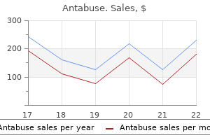
Antabuse dosages: 500 mg, 250 mg
Antabuse packs: 30 pills, 60 pills, 90 pills, 120 pills, 180 pills, 270 pills, 360 pills

Buy cheap antabuse 500 mg on line
Damage is more probably to happen when the cranium is "rebounding" from the impact and the vessels are experiencing tensile strain useless id symptoms antabuse 500 mg order online. For cases of cranium fracture, the localized pressure from the skull contacting the cortical surface offers a way for the vascular damage famous within the pia and cortex symptoms ibs buy 500 mg antabuse overnight delivery. Contrecoup Contusions Two phenomena have been attributed to the pathogenesis of countercoup contusions: cavitation results and inertial loading. Of the two, the extra likely mechanism of contrecoup harm is translational or angular head movement. On impression, the brain moves toward the influence web site and creates an space of adverse pressure directly reverse the loading level. This negative strain could in flip trigger injury by exceeding the tensile strength of water or, alternatively, cause small gas bubbles to seem within the parenchyma. The return to regular or constructive stress will cause the small bubbles to break down and is termed cavitation. Instead, it appears that the areas of vascular disruption and cortical harm and contrecoup areas are due primarily to acceleration effects and may end up from either translation or angular head motions. Each head motion, notably angular motion, is able to producing tensile pressure throughout the mind. If the tensile strain is greater than the vascular tolerance in a given region, contusion occurs. The term "contrecoup" can subsequently be thought-about deceptive as a result of the important mechanism is most frequently acceleration and not the contact effects from impression. In conditions during which the pinnacle experiences impulsive loading, contrecoup contusions happen solely because of the pressure generated in the cortical brain region during the acceleration period. Strain concentrations could occur in specific regions of the mind because of geometric results and are accountable partially for the high incidence of frontal and temporal lobe contusions observed clinically. Although global skull deformation caused by influence could create tensile stress and contusion damage in regions remote from the influence, the predominant mechanism of contrecoup contusions is rotational acceleration. Because of their macroscopic and easily identifiable nature, contusions are periodically used to characterize the biomechanical input to the head. However, several points deserve point out when using contusions as a software on this method. The discussion within the previous paragraphs highlights the importance of inertial loading producing the pattern of contrecoup damage versus the function of native contact effects. Thus, coup and contrecoup injuries, though differing only slightly in name, arise from basically completely different mechanisms. In reality, essentially the most frequently occurring contusions of the temporal and frontal poles are contrecoup in nearly every occasion, regardless of the impression site. Rather, the relative proportion of coup versus contrecoup contusions depends solely on the response of the head to impact. Because of its pathogenesis, the acceleration of the top brought on by a concentrated blow to the head has led to the proposal that acceleration causes coup contusions. Local cranium deformation produces a noticeable coup contusion, and the dearth of substantial head motion produces slight or very restricted contrecoup contusion. Conversely, a softer or larger influence object is commonly seen in instances of deceleration injury, corresponding to in a car accident sufferer, in whom the injuries are caused less by native damage beneath the point of impression as a end result of a proportional improve in vitality is utilized in setting the pinnacle into movement or stopping it from transferring. In this case, a big contrecoup area happens and the coup contusion is smaller or nonexistent due to the delicate, extensive padded surfaces inside modern automobiles. Subdural Hematoma Three sorts of acute subdural hematoma are discovered clinically. The first two are related to contusion and laceration and are typically referred to as difficult subdural hematomas. Complicated subdural hematomas result from the contact or acceleration effects that trigger the first lesion. The third sort of subdural hematoma is the most typical form of vascular disruption; it involves tearing of parasagittal bridging veins positioned along the interhemispheric fissure and sagittal sinus. Based on their superficial location, parasagittal bridging veins are vulnerable to damage throughout short-duration angular acceleration of the head. Common conditions by which this acceleration situation occurs embody falls during which the pinnacle strikes a broad, onerous surface and assaults by which a majority of the impact energy is used to set the top in movement. Under these loading conditions, the pressure inside the mind is concentrated along the outer margins where the parasagittal bridging veins reside. In the latter case, the brain is believed to strike or be pulled away from adjacent, comparatively motionless buildings, a mechanism that ends in focal or compressive pressure, respectively. Alternatively, intermediate contusions of the cingulate gyrus could be because of interactions with the falx, and equally, those of the inferomedial temporal lobe may finish up from involvement with the tentorium of the petrous ridge. Diffuse Brain Injury Cerebral Concussion All gradations of concussion (transient reversible neurological dysfunction as a outcome of trauma) are produced totally by inertial loading and not from contact phenomena effects. Angular rotational head motion causes the deeper structures within the brain to deform and leads to the classic widespread disruption of mind function that underlies concussion. For a concussive injury, most of the pressure is insufficient to cause structural injury. Instead, damage to the structures may be both partially or utterly reversible, relying on the severity of the inertial loading. The precise location of the functional derangement in a concussion continues to be debated. It remains uncertain whether or not the consequences of angular acceleration are principally seen in the brainstem, the cerebral hemispheres, or each regions. Intracerebral Hematoma Large traumatic intracerebral hematomas are uncommon and are most often related to intensive cortical contusions. They can subsequently be considered contusions in which bigger, deeper vessels have been disrupted. Accordingly, these hemorrhages are considered to be as a result of inertial or head movement results and are therefore not associated to contact phenomena. Tissue tear hemorrhages are sometimes numerous, small, and positioned parasagittally and within the central third of the mind. Their areas are characteristically within the superior medial frontoparietal white matter, corpus callosum, centrum semiovale, periventricular white and grey matter ("gliding contusions"), inner capsule, and basal ganglia. In the brainstem, they appear in dorsal area of the midbrain and upper pons ("dorsolateral quadrant brainstem contusions"). Several factors contribute to those multiple foci of damage, together with the presence of intracranial partitioning membranes, the geometric irregularities of the cranium, and the airplane of motion experienced by the pinnacle. Diffuse Axonal Injury Axonal damage seems to be one of the two most important pathologic substrates producing extended traumatic coma not attributable to mass lesions and, like cerebral concussion, is brought on only by angular rotational acceleration and never by contact phenomena (the second pathologic substrate being ischemic/ hypoxic neuronal damage). Critical elements in estimating the amount and extent of axonal injury include the magnitude, length, and onset rate of the angular acceleration, in addition to the path of movement and the role of the intracranial membranes. The path that the pinnacle moves performs an essential role in the quantity and distribution of axonal harm in a given state of affairs. For equivalent levels of angular acceleration, the brain is most weak to axonal harm if it is moved laterally.
Best antabuse 500 mg
Maxillary Fracture Patterns Maxillary fractures are nonetheless most frequently classified based on the unique description by Le Fort medicine 20th century 250 mg antabuse effective. Head Neck Le Fort I-a horizontal fracture via the piriform aperture, the zygomatic buttresses, and the pterygoid plates symptoms of hiv generic 500 mg antabuse visa. The medical indicators rely upon the degree of displacement and on whether the fracture extends to the orbits and nasal bone. There may be bruising, swelling, and elongation of the midface, mobility of the midface, and malocclusion. In an edentulous patient with only minor radiologic proof of displacement and in whom the fracture appears to be steady with mastication. The pretrauma occlusion is set and an acrylic chew wafer constructed on the dental solid. This is used perioperatively and often postoperatively to keep up the right occlusion. Because upper jaw fractures may compromise both the oral and nasal cavities, preoperative airway management wants careful planning. After cautious disimpaction and mobilization of the fractures, an acrylic occlusal wafer is wired to the higher jaw. The relationship of the upper jaw to the midface buttresses is established with the mandibular condyles properly seated within the glenoid fossae. If any of those buttresses is comminuted, vertical height may be maintained with bone grafting. After all 4 buttresses are stabilized with miniplates or bone grafting, the intermaxillary fixation could additionally be released and the success of the reduction confirmed by seeing the lower enamel have interaction cleanly into the wafer. The fracture is then supported with one or two very light elastic bands between the higher and decrease jaws. The occlusal complicated, which consists of the higher and lower jaws united, is then brought as much as the stabilized upper midface, and the final fixation, with or with out bone grafting, is carried out alongside the traces of the buttresses. The acrylic occlusal wafer may be left on the upper jaw and the mandible permitted to have interaction within the dental sides on the inferior aspect of the wafer for a variety of days. Light rubber band traction may be placed between the higher and decrease jaws in the premolar area after the first 24 hours to inhibit contraction of the medial pterygoid muscular tissues during the earlier levels of mobilization. Patients are nursed while sitting up as soon as potential to scale back facial swelling, and jaw mobilization is commenced nearly instantly. Repair requires repositioning present bone, changing severely comminuted or misplaced bone with major autogenous bone grafts, and replacing or increasing craniofacial gentle tissues to the pretraumatic state. The first step in management is to revive midface projection and width in relation to the cranial base and mandible. Gruss and associates grouped these complex injuries into three broad anatomic divisions43: 1. Central-a central impact to the frontonasal area that splits the central facial buttress from the anterior cranium base. B, After reconstruction of the panfacial fracture in A, by mandibularplating,zygomaticplating,bonegrafting of the comminuted area of the left zygoma and maxilla, plating alongside the buttresses of the maxilla,andabonegrafttothenasaldorsum. A B Fractures could prolong into the frontal bone, orbital roof, midface, and central mandible. Lateral-a lateral impact damaging the lateral facial buttress (frontozygomaticomaxillary) and increasing into the greater wing of the sphenoid, temporal, and parietal bones. Combined central and lateral-a major impact inflicting extensive comminution of the central and lateral elements, often with sagittal fracturing of the midface and mandible and with condylar fractures. The cranio-orbital interface is the junction between the anterior cranial fossa and frontal sinus above and the orbits and nasoethmoid area below. A safe interface between the contents of the anterior cranial fossa and the nasal cavity is essential for avoiding intracranial infection. Extension of cranial base fractures via the sphenoid bone could injure the neurovascular buildings of the orbital apex, superior orbital fissure, and cavernous sinus. Damage to the orbital roof and lateral partitions may have an effect on the projection or motility of the eye (or both) or cause orbital pulsation. The zygomatic arch and the lateral orbital wall most precisely relate the orbit and midface to the cranial base above. Correct steady arch position determines the lateral midface projection and midfacial width. Reconstruction of the maxilla in its correct place between the zygomas will complete the midfacial arch and central midfacial projection with a proper relationship to the mandible. The mandibular condyles demarcate the posterolateral restrict of the craniofacial interface. The condyles are regularly injured with panfacial fractures and must be carefully restored to reestablish the vertical dimension of the face. The midface must be related to the intact mandibular arch at the occlusal degree as a result of the mandibular arch determines facial top and, to a lesser extent, central facial projection. Clinical Assessment After quick resuscitation as set out earlier, neurological and ophthalmologic assessments have to be undertaken. There may be marked bruising and swelling, deformity, and abnormal movement of the facial skeleton. In very advanced instances, nylon models generated from the scan are valuable in planning the reconstructive surgery. Treatment Complex craniofacial fractures ideally require definitive correction of the bony injuries inside the first 5 to 7 days. As famous earlier, some purpose for primary fixation within 12 to 48 hours of injury, however this early time-frame may not allow adequate preoperative radiologic, ophthalmologic, and dental evaluation, particularly when the affected person is comatose or poorly cooperative. During this early section, delicate tissue swelling makes operative publicity and assessment of facial projection and symmetry troublesome. When early acute neurosurgical intervention is important for intracranial clots or compound calvarial fractures, the scalp incisions should be designed to permit later publicity and stabilization of the craniofacial fractures. Because fixation of a cellular face requires an intact fronto-orbital bar, a frontal bone flap elevated to show the anterior cranial fossa should be positioned to take care of the secure superior points for fixation. When treating a depressed fracture of the temporal area, it may be possible to stabilize fractures extending into the lateral orbital wall and greater wing of the sphenoid. Indeed, delayed restore of an orbital fracture might risk displacing a beforehand corrected cranial element and causing intracranial bleeding or dural harm. The key steps on this sequence are correct positioning and fixation of the zygomatic arches. If the mandibular arch is disrupted, particularly if it is foreshortened because of fracture or fracture-dislocation of the condylar processes, it must be repaired initially to establish a solid foundation and posterior facial top. Once the mandible is rigidly restored, the midface may be disimpacted and positioned within the predicted occlusion. Repair subsequently proceeds from above down to meet the already fastened maxillary-mandibular section. Miniplate fixation or bone grafting is used to reconstitute the four anterior buttresses.
500 mg antabuse cheap with mastercard
Pha rma codyna mi cs /Ki neti cs Ha l f-l i fe el i mi na ti on: 10-20 hours Pha rma cothera py Pea rl s Ca rbi noxa mi ne ma l ea the i s ~71% ca rbi noxa mi ne in treatment 1-3 antabuse 500 mg order line. Ca rbo-Ta x (Adenoca rci noma) Lexi -Drugs Onl i ne Jump To Fi el d (Sel ect Fi el d Na me) Pha rma col ogi c Ca tegoryChemothera py Regi males, Adenoca rci noma (Unknown Pri ma ry) Us e: La bel ed Indi ca ti ons Adenoca rci noma, unknown pri ma ry Regi men Us eAdenoca rci noma, unknown pri ma ry Index Terms Pa cl i ta xel -Ca rbopl a ti n (Adenoca rci noma) Regi males Pa cl i ta xel: I medicine 93 7338 250 mg antabuse discount with mastercard. Ca l cul a ti ons Body Surfa ce Area: Adul ts Body Surfa ce Area: Pedi a tri cs Ca l vert Formul a Admi ni s tra ti on: I. Recons ti tuted ca rbopl a ti n 10 mg/mL s houl d be additional di l uted to a fi na l concentra ti on of zero. Compatibility when admixed: Compatible: Ci s pl a ti n, etopos i de, fl oxuri di ne, i fos fa mi de, i fos fa mi de wi th etopos i de, pa cl i ta xel. Ma l e/fema l e: Cons ul t pres cri ber for i ns tructi on on a ppropri a the contra cepti ve mea s ures. Concerns related to adverse results: � Hypertens i on: Increa s ed bl ood pres s ure ma y be obs erved wi th trea tment. Boxed Warning] Potent oxytocic agent; use with strict adherence to recommended dosing. Moni tor bl ood pres s ure, thera peuti c effecti venes s, a nd a dvers e rea cti ons. Dos i ng: Pedi a tri cRefer to i ndi vi dua l protocol s: Chi l dren (unl a bel ed us e): I. Acute l ung i njury tends to occur 1-3 months fol l owi ng ca rmus ti ne i nfus i on. Oxi di zed regenera ted cel l ul os e (Surgi cel ) ma y be pl a ced over the wa fer to s ecure; i rri ga the ca vi ty pri or to cl os ure. Ma y ca us e bl eedi ng (due to thrombocytopeni a) or i nfecti ons (due to neutropeni a); moni tor cl os el y. Boxed Warnings]: Dose-related pulmonary toxicity might occur; patients receiving cumulative doses >1400 mg/m2 are at higher risk. Delayed onset of pulmonary fibrosis has occurred up to 17 years after treatment in youngsters (1-16 years) who acquired carmustine in cumulative doses ranging from 770-1800 mg/m2 mixed with cranial radiotherapy for intracranial tumors. Advers e Rea cti ons >10%: Ca rdi ova s cul a r: Hypotens i on (wi th hello gh-dos e I. Ca ps ul es ma y be opened a nd s pri nkl ed on a ppl es a uce for i mmedi a the us. Risk C: Monitor therapy Sel ecti ve Serotoni n Reupta ke Inhi bi tors: Ma y enha nce the bra dyca rdi c impact of Beta -Bl ockers. Risk X: Avoid combination Etha nol /Nutri ti on/Herb Intera cti ons Food: Food decrea s es ra the however not extent of a bs orpti on. Admi ni s tra ti on wi th meals mi ni mi zes ri s ks of orthos ta ti c hypotens i on. Denta l Hea l th: Effects on Denta l Trea tmentKey a dvers e event(s) rel a ted to denta l trea tment: Pos tura l hypotens i on. Pros pecti ve Ra ndomi zed Eva l ua ti on of Ca rvedi l ol on Symptoms a nd Exerci s e," Circulation, 1996, 94(11):2793-9. Dos i ng: Pedi a tri c Aspergillosis, candidiasis, empiric therapy: Chi l dren >3 months to 17 yea rs: I. Recons ti tuted s ol uti on ma y be s tored a t 25�C (77�F) for 1 hour pri or to prepa ra ti on of i nfus i on s ol uti on. As s es s thera peuti c effecti venes s (a ccordi ng to purpos e for us e) a nd moni tor cl os el y for a dvers e rea cti ons. Moni tori ng: La b Tes ts Li ver functi on Pa ti ent Educa ti onThi s medi ca ti on ca n onl y be a dmi ni s tered by i nfus i on. Di eta ry Cons i dera ti ons Ca ps ul e, chewa bl e ta bl et, a nd s us pens i on ma y be ta ken wi th or wi thout food. The ma nufa cturer recommends tha t ca uti on be exerci s ed when a dmi ni s teri ng cefa cl or to nurs i ng ladies. The ma nufa cturer recommends tha t ca uti on be exerci s ed when a dmi ni s teri ng cefa droxi l to nurs i ng girls. Compatibility when admixed: Compatible: Aztreona m, cl i nda myci n, fa moti di ne, fl ucona zol e, l i nezol i d, meperi di ne, metroni da zol e, metroni da zol e wi th s odi um bi ca rbona te, vera pa mi l. Subs equent dos es (300 mg or 7 mg/kg/dos e) s houl d be a dmi ni s tered every other da y. Moni tori ng: La b Tes ts Perform cul ture a nd s ens i ti vi ty s tudi es pri or to i ni ti a ti ng drug thera py; rena l functi on. Pha rma codyna mi cs /Ki neti cs Di s tri buti on: Vd: Chi l dren 6 months to 12 yea rs: 0. The ma nufa cturer recommends ca uti on when us i ng cefdi toren duri ng brea s t-feedi ng. Moni tori ng: La b Tes ts Perform cul ture a nd s ens i ti vi ty s tudi es pri or to i ni ti a ti ng drug thera py; rena l functi on Pa ti ent Educa ti onDo not ta ke a ny new medi ca ti on duri ng thera py wi thout cons ul ti ng pres cri ber. V: 2 g every eight hours for 7 da ys or unti l the neutropeni a res ol ves Intra-abdominal infections, difficult: I. Community-acquired (including pseudomonal): 1-2 g each 12 hours for 10 da ys Septic lateral/cavernous sinus thrombosis (unlabeled use): I. The ma nufa cturer recommends tha t ca uti on be exerci s ed when a dmi ni s teri ng cefepi me to nurs i ng women. Dos i ng: Pedi a tri c Susceptible infections: Ora l: Chi l dren 6 months: 8 mg/kg/da y di vi ded every 12-24 hours Chi l dren >50 kg or >12 yea rs: Refer to a dul t dos i ng. Risk C: Monitor therapy Etha nol /Nutri ti on/Herb Intera cti ons Food: Del a ys cefi xi me a bs orpti on. Dos i ng: Pedi a tri c Infa nts a nd Chi l dren 1 month to 12 yea rs: Susceptible infections: I. Dos i ng: Rena l Impa i rment Cl cr 10-50 mL/mi nute: Admi ni s ter every 8-12 hours. Incompatible: Al l opuri nol, fi l gra s ti m, fl ucona zol e, gemci ta bi ne, heta s ta rch, penta mi di ne. Compatibility when admixed: Compatible: Cl i nda myci n, metroni da zol e, vera pa mi l. The ma nufa cturer recommends tha t ca uti on be exerci s ed when a dmi ni s teri ng cefota xi me to nurs i ng women. Conti nuous a rteri ovenous or venovenous hemodi a fi l tra ti on effects: Admi ni s ter 750 mg every 12 hours Ca l cul a ti ons Crea ti ni ne Cl ea ra nce: Adul ts Crea ti ni ne Cl ea ra nce: Pedi a tri cs Admi ni s tra ti on: I. The ma nufa cturer recommends ca uti on when gi vi ng cefoteta n to a brea s t-feedi ng mother. Injecti on, powder for recons ti tuti on: 1 g, 2 g [conta i ns s odi um eighty mg/g (3.
Antabuse 250 mg trusted
Modifications of the Pterional Approach In some instances, the normal pterional craniotomy could be modified to improve the surgical exposure, restrict mind retraction, and improve entry to tough lesions medications given during dialysis generic antabuse 500 mg fast delivery. The superolateral orbit and the zygomatic arch as a unit is eliminated, separate from the bone flap medicine synonym cheap antabuse 500 mg with visa. This increases the publicity associated with the pterional approach (A) and provides a (B) completely different trajectory (more upward). An extended orbital-zygomatic method, during which a subtemporal decompression is also performed, can improve the inferior exposure. These approaches are discussed in more detail in the part on Posterior Circulation aneurysms. The affected person is placed supine with the pinnacle flexed about 20 levels but not to compromise venous drainage. The head is positioned straight, or slightly angled towards the contralateral facet. A bicoronal pores and skin incision behind the hairline or U-shaped frontal incision, with the base of the U simply on the contralateral facet of the sagittal sinus may be used. The bone flap is rectangular in form and may lengthen past midline; this helps cut back frontal lobe retraction as a end result of the falx can be retracted. Frameless stereotaxy may be a helpful adjunct to make certain that the bone opening is anterior enough to permit proximal management. A, the identical old craniotomy for pericallosal artery aneurysms is located anterior to the coronal suture. The medial bone edge is a minimal of at the midline to reveal the superior sagittal sinus. B, the aneurysm is exposed via an interhemispheric method with a retractor on the falx for visualization. In a affected person with a quantity of aneurysms we avoid simultaneous pterional and parasagittal craniotomies. In this section we evaluation the varied surgical approaches in accordance with the three main vascular territories. Dissections and fusiform aneurysms are more widespread within the posterior than in the anterior circulation. Many posterior circulation aneurysms are tough to access because of the deep midline location of the vertebrobasilar system, confinement by the clivus and petrous pyramids, and the shut relationship to the cranial nerves. Consequently, as endovascular techniques have advanced, direct surgery on posterior circulation aneurysms nows much less frequent. There are several surgical approaches to these lesions outlined by the uncovered vascular territory (basilar apex, basilar trunk, and vertebral trunk) and surgical trajectory (anterosuperior, lateral, and posteroinferior; Table 365-3). There are several surgical approaches to these aneurysms (Table 365-4); the prolonged orbitozygomatic approach provides the best publicity and adaptability of trajectories. Careful choice of an strategy is critical to surgical success and is, in massive part, influenced by aneurysm morphology together with: (1) aneurysm web site and dimension, (2) exact origin of the sac, (3) fundus projection and dimension, (4) clival degree of the bifurcation, (5) distance from the sagittal midline, and (6) distance from the clivus. In common, extracranial modification increases access and reduces retraction, whereas intracranial modification supplies access to the instant neighborhood. When the bifurcation is situated more than 1 cm below the level of the posterior clinoids, its view typically is obscured when using a pterional transsylvian strategy and so these lesions could additionally be better approached using a subtemporal trajectory, modified if essential with a medial petrosectomy or division of the tentorium to succeed in down the clivus. Lesions on the stage of the posterior clinoid and as a lot as 1cm above the clinoids may be approached utilizing a subtemporal or transsylvian strategy. However, the higher the bifurcation is relative to the posterior clinoid, greater temporal lobe retraction is required. Instead, the craniotomy requires modification corresponding to elimination of the zygoma or fronto-orbital bone (orbitozygomatic approach). Subtemporal the subtemporal strategy proceeds from a lateral trajectory under the temporal lobe and along the middle fossa flooring. The space behind the aneurysm, including the perforators, whose preservation is essential, usually is seen best from this strategy. There are several disadvantages to the subtemporal method: (1) the operating field is small; (2) extra temporal lobe retraction could additionally be needed; (3) the ipsilateral P1 lies between the surgeon and the aneurysm, which can limit dissection or clip utility; (4) the aneurysm, significantly when large, needs to be retracted to see the alternative P1; and (5) a high-lying bifurcation could additionally be troublesome to approach. The affected person is positioned in the lateral decubitus or in the supine place with a shoulder roll. The head is rotated till the midline plane (superior sagittal sinus) is parallel to the floor, and the vertex is angled 15 to 20 degrees downward to realize a line of sight parallel to the floor of the center fossa. One of two incisions may be used: a 7- to 10-cm linear incision that extends up from a point 1 cm anterior to the tragus at the zygomatic arch or a query mark that starts just anterior to the tragus and curves above the ear to the superior temporal line. A 4- � 4-cm craniotomy is made and temporal squamosal bone removed inferiorly with a bur until flush with the middle fossa ground. Under the working microscope, a self-retaining retractor is positioned to raise the temporal lobe and expose the tentorial incisura. Dissection then is directed between the medial temporal lobe and the tentorial edge. Temporal bone is removed inferiorly to make the cranial publicity flush with the middle fossa floor (crosshatching). B, Microsurgical anatomy of the tentorial incisura as soon as the temporal lobe is elevated. The fundamental approach is just like a pterional craniotomy for anterior circulation aneurysms. A frontotemporal craniotomy is made that extends medially to the supraorbital nerve foramen and inferiorly to the floor of the anterior cranial fossa. This extensive bone removal helps present a flat anterior fossa and anterior middle fossa trajectory. It may be essential to divide temporal bridging veins to mobilize the temporal pole; posterior temporal lobe retraction often is better tolerated than lifting the lobe up. The lateral retrocarotid area provides the least restricted angle of clip utility. Access via the medial retrocarotid hall is beneficial when the basilar bifurcation is excessive, the P1 phase is short, or the PcomA is inflexible and runs concave lateral. Often, multiple routes are needed or totally different routes are used concurrently to cross an instrument, suction, or clip applier. During aneurysm dissection, the microscope is moved to place the third nerve within the middle of subject. Orbitozygomatic-Pterional Approach the orbitozygomatic strategy adds two steps to the pterional method: (1) gentle tissue dissection to reveal the orbitozygomatic unit (the orbital rim, orbital roof, lateral orbital wall, and zygomatic arch), and (2) osteotomies to free it. This method is beneficial for a high bifurcation and offers a extra anterior trajectory, a higher view above the posterior clinoid course of, and larger area within the operative hall than a normal pterional craniotomy. The zygoma and orbital rim are ensheathed in two layers of the temporalis fascia that can be elevated via an interfascial dissection that peels aside the layers or by a subfascial dissection.
Cheap antabuse 500 mg mastercard
Taken together, the pure historical past of the patient and the natural historical past of the particular aneurysm have to be thought-about in evaluating the overall method to treatment medicine 7 years nigeria discount antabuse 250 mg without prescription. For example, the treatment of a small basilar tip aneurysm in a 40-year-old and in an 80-year-old will differ regardless of the similarity within the lesion symptoms quadriceps tendonitis antabuse 250 mg online buy cheap. Patient concerns and the pure historical past of cerebral aneurysms are comprehensively reviewed in Chapters 354 and 360. As outlined later, there are similarities, however mainly significant differences in affected person care choices. Tsutsumi and coworkers2 adopted 115 sufferers for almost 9 years after their unruptured aneurysm had been clipped and found only one affected person (0. A combination of imaging techniques often is critical, particularly for more advanced aneurysms; this permits correct preoperative planning and a full understanding of the related anatomy and these research will usually be crucial in determining timing. Most case series3-9 reveal that the morbidity and mortality related to surgical therapy of unruptured aneurysms is very low; about 5% of sufferers die or are disabled. For example, King and associates14 carried out a meta-analysis of 28 scientific series containing 733 sufferers; overall, four. Similarly, Raaymakers and coworkers15 carried out a meta-analysis from a Medline search between 1966 and 1996 and recognized 61 studies involving 2460 sufferers. It must be acknowledged that a lot of the info on outcomes of aneurysm surgery additionally come from high quantity facilities and single-center studies, and publication bias particularly must be considered when making use of the reported dangers of aneurysm surgical procedure to a broader population. Similarly, Solomon and coworkers7 noticed a positive end result in 83% of sufferers who underwent surgical repair of large unruptured aneurysms. By distinction, 99% of patients who had aneurysms lower than 10 mm in diameter skilled good outcomes. Repair of unruptured posterior circulation aneurysms, particularly large basilar bifurcation aneurysms, is related to increased surgical danger. By distinction, the morbidity and mortality for large anterior circulation unruptured aneurysms was 13%. Reasonable surgical results may be expected with nongiant unruptured posterior circulation aneurysms. Calcification within the aneurysm neck typically is related to poor consequence, partially, as a end result of many of those lesions are massive or large aneurysms. In addition, multiple clips frequently are necessary to occlude the aneurysm, leading to an elevated incidence of cerebral embolism. Calcification may be eliminated by endarterectomy; despite this, surgical outcomes remain poor as a result of the remaining wall for clip placement is usually very friable and skinny. Hypothermic circulatory arrest ought to be thought-about in the reconstruction of some heavily calcified aneurysms. There are a quantity of patient-related components including advanced age, ischemic cerebrovascular disease, and medical situations such as diabetes mellitus that improve the risk of unruptured aneurysm surgical procedure. In 1994 and 200922,23 the American Heart Association revealed guidelines for the therapy of unruptured intracranial aneurysms. Since then a number of studies have improved our understanding of the natural historical past and surgical threat and alternate therapies for unruptured aneurysms have developed. Microsurgical, endovascular, and different methods similar to vessel occlusion with or with out bypass or no remedy apart from remark and medical follow-up ought to be thought-about, specific to the patient and the aneurysm. B,Histogram illustrating that grade, which is set by adding the purpose value for every factor, is strongly associated with surgical consequence. Predicting end result following surgical remedy of unruptured intracranial aneurysms: A proposed grading system. This scale was derived from a retrospective analysis of 172 sufferers who underwent therapy of unruptured aneurysms after which validated prospectively in 50 sufferers. Garrett and associates24 lately performed a complete literature review (10,545 patients) and using mathematical models created a web-based open entry program to quantify the benefit of surgical procedure on unruptured aneurysms in accordance with age, size, and placement. Small basilar and supraclinoid aneurysms (5 mm) solely had an operative benefit in young patients (40 years). By distinction, bigger aneurysms (10 to fifteen mm) had an operative profit for a wider age range. There was no age and size mixture that provided an operative profit for cavernous aneurysms. However, because few asymptomatic aneurysms truly rupture and because all aneurysm remedy carries some threat, the management of patients with an unruptured aneurysm stays controversial. These recommendations depend upon collaboration between a highly experienced cerebrovascular group of microneurosurgeons and endovascular neurosurgeons at a tertiary medical heart with a high case quantity. In addition sufferers who smoke and are hypertensive could additionally be thought of for remedy when the aneurysm is small. Consequently recommendation of serial imaging is considerably controversial (we get hold of serial noninvasive imaging each 6 months). The position of endovascular techniques for unruptured aneurysms and tips on how to determine which sufferers ought to bear open microsurgery or an endovascular procedure is mentioned later within the chapter. Untreated, between 20% and 30% of aneurysms rerupture throughout the first 30 days and then at a price of approximately 3% to 5% per 12 months. Aneurysm rebleeding may be lowered by antifibrinolytic administration, however can only be prevented by direct obliteration using surgical or endovascular strategies. There are two important questions: when ought to surgery be carried out and which patients ought to endure surgical or endovascular aneurysm occlusion Timing of Aneurysm Obliteration Early surgery eradicates the danger of rebleeding and appears to be associated with improved consequence. For instance, among the 722 sufferers handled at 27 North American centers within the International Cooperative Study on the Timing of Aneurysm Surgery, a prospective epidemiologic, but nonrandomized examine discovered early surgery significantly improved consequence. Brilstra and associates36 examined how effective aneurysm clipping was at decreasing poor outcome associated with rebleeding: a threat discount of 19% was observed in patients who had surgery rather than conservative management. For each affected person with a ruptured aneurysm, there are a quantity of components, together with aneurysm-related, hemorrhage-related, and patient-related factors, that influence whether or not to function acutely or in a delayed trend. Advanced age is related to poor end result; however, this could not exclude the affected person from early surgical procedure, partly as a outcome of elderly patients usually tend to have intracerebral hematomas and a decreased cerebrovascular reserve that increases their threat for delayed ischemia. Delayed surgery could additionally be preferable for complicated lesions similar to large aneurysms or those the place extended durations of short-term occlusion are expected to achieve aneurysm occlusion. Nonrandomized studies that describe concurrent cohorts or historical controls and enormous medical series35,42-48 have observed an inclination for patients undergoing early surgery to experience better outcomes. The timing of aneurysm obliteration for posterior circulation aneurysms is much less nicely outlined. In addition, as a end result of many of those patients now bear endovascular procedures, the question may be much less related. Most info that favored delayed surgery got here from specialised referral facilities. Fifty percent of patients scheduled for late surgery (n = 26) made an excellent restoration, whereas 72% scheduled for early surgery (n = 23) made a good recovery. When surgery is indicated rather than endovascular occlusion, we recommend early surgery for ruptured posterior circulation aneurysms apart from those lesions which will current technical problem, corresponding to giant posterior-oriented basilar bifurcation aneurysms. Earlier aneurysm occlusion utilizing surgical or endovascular techniques has reduced the impact of rebleeding on consequence.
250 mg antabuse discount with mastercard
Ratings are generally based both on behavioral signs and on confusion and cognitive impairment medicine 8 soundcloud buy antabuse 250 mg amex. Rating the severity of delirium over time could additionally be helpful for monitoring the effect of an intervention or plotting the course of a delirium over time symptoms underactive thyroid 500 mg antabuse trusted. These scales have also been used to make the prognosis of delirium by considering sufferers with scores above a specified cutoff to have the diagnosis. Laboratory checks Several laboratory evaluations have been investigated for possible use in evaluating delirium. Among the experimental laboratory checks which were investigated to be used in delirium, those that seem to indicate some promise embody mind imaging (46, 47) and measures of serum anticholinergic activity (48). First, the cornerstone of therapy, psychiatric management, is defined and its elements are described. Treatment of the delirium itself entails a set of environmental and supportive interventions and particular pharmacologic remedies. Environmental manipulations are typically designed to help reorient the patient and modulate the diploma of stimulation. Supportive measures are designed to provide patient, household, and associates with both reassurance and training concerning the nature, temporal course, and sequelae of delirium. A psychologically knowledgeable understanding of the affected person and the household may facilitate these duties. In many circumstances, the psychiatrist might be part of, or a advisor to, a multidisciplinary team and will encourage the administration of the full range of needed remedies. Coordinate with other physicians caring for the affected person Delirium frequently heralds a medical emergency, and patients are usually managed in an acute-care hospital setting. For some sufferers with milder symptoms, as soon as the etiology of delirium has been identified and general medical management has begun, psychiatric and general medical administration can happen in an alternate setting. The psychiatrist is usually asked to seek the assistance of when a patient develops delirium on a basic medical or surgical unit in the hospital; however, delirium may current as an emergency in both the psychiatric outpatient or inpatient setting. The appropriate treatment of delirium entails interventions to search for and proper underlying causes, in addition to relieve current signs. Joint and coordinated administration of the patient with delirium by the psychiatrist and internist, neurologist, or different major care or specialty physicians will incessantly assist guarantee acceptable comprehensive evaluation and care. Identify the etiology An essential principle in the psychiatric administration of delirium is the identification and correction of the etiologic elements. Appropriate laboratory and radiological investigations such as those listed in Table three may be essential to find out the underlying cause(s) of delirium. The selection of particular checks to be undertaken will rely upon the outcomes of the clinical analysis. Initiate interventions for acute circumstances A patient with delirium might have life-threatening general medical situations that demand therapeutic intervention even before a particular or definitive etiology is determined. In addition to making sure that diagnostic tests important to identifying the cause for delirium are ordered, when appearing as a consultant, the psychiatrist should elevate the extent of consciousness of the general medical staff concerning the potential morbidity and mortality related to delirium. Treatment of Patients With Delirium 17 Copyright 2010, American Psychiatric Association. Assessment of the Patient With Delirium Domain Measure Physical standing History Physical and neurological examinations Review of important indicators and anesthesia document if postoperative Review of general medical information Careful evaluate of medicines and correlation with behavioral adjustments Interview Cognitive tests. Provide other disorder-specific remedy the objective of analysis is to find reversible causes of delirium and forestall issues via immediate remedy of those particular disorders. One must give a high precedence to identifying and treating such problems as hypoglycemia, hypoxia or anoxia, hyperthermia, hypertension, thiamine deficiency, withdrawal states, and anticholinergic-induced or other substanceinduced delirium. Examples of particular reversible causes of delirium and treatments for these disorders seem in Table four. Monitor and guarantee safety Behavioral disturbances, cognitive deficits, and other manifestations of delirium might endanger sufferers or others. Psychiatrists must assess the suicidality and violence potential of sufferers and implement or advocate interventions to attenuate these risks. Suicidal behaviors are sometimes inadvertent in delirium and happen in the context of cognitive impairment and/or in response to hallucinations or delusions. Examples of Reversible Causes of Delirium and Their Treatments Condition Treatment Hypoglycemia or delirium of unknown etiology the place hypoglycemia is suspected Hypoxia or anoxia. Immediate oxygen Rapid cooling Prompt antihypertensive therapy Appropriate pharmacologic intervention Thiamine, intravenous glucose, magnesium, phosphate, and other B vitamins, together with folate Thiamine hydrochloride, 100 mg i. Additional guidelines may apply in some jurisdictions, and the psychiatrist ought to become conversant in relevant regulations and institutional policies (52). The symptoms and behavioral manifestations of delirium can fluctuate quickly, and common monitoring will permit for the adjustment of therapy methods. Important behavioral issues that must be addressed include depression, suicidal ideation or habits, hallucinations, delusions, aggressive habits, agitation, anxiety, disinhibition, affective lability, cognitive deficits, and sleep disturbances. It is helpful to report serial assessments of psychological standing and symptoms over time, as these may point out the effectiveness of interventions and new or worsening medical conditions. This understanding may be based mostly on prior acquaintance with the affected person, current interviews or interplay with the patient or household, and/or history from the family. Treatment of Patients With Delirium 19 Copyright 2010, American Psychiatric Association. Understanding the underlying affect, concerns, and premorbid persona of the patient is incessantly helpful in maintaining a supportive alliance. A solid alliance with the household can be desirable, as relations are a important supply of potential support for sufferers and knowledge on patients who may be unable to give dependable histories. Establishing robust alliances with the multiple clinicians and caregivers frequently involved within the care of delirious medically sick patients is also crucial. Educate affected person and family concerning the illness Educating sufferers and families relating to delirium, its etiology, and its course is a crucial position for psychiatrists involved in the care of sufferers with delirium. Patients might range of their capacity to understand their situation; however, offering reassurance that delirium is normally short-term and that the signs are a part of a medical condition may be extremely beneficial to both sufferers and their households. Specific educational and supportive interventions are mentioned in more element in the following paragraphs. Nursing staff make frequent observations of patients over time, which places them in a wonderful position to detect the onset and monitor the course of delirium. Education of nursing workers on each shift concerning the clinical features and course of delirium could be an essential task for psychiatrists. In such cases, offering training to different physicians concerning the underlying physiological etiologies of delirium may be an important task for the psychiatrist. Some patients progressively or abruptly lose all apparent recall of the delirious experience, whereas others have vivid, frightening recollections. Explanations relating to delirium, its etiology, and its course must be reiterated. Supportive interventions which may be a standard a part of psychiatric administration following a traumatic expertise should be used for those with distressing postdelirium symptoms. Psychotherapy centered on working via the expertise of the delirium may, at times, be essential to resolve anxiety, guilt, anger, melancholy, or other emotional states. Environmental interventions Management of delirium features a specific array of interventions by nursing, psychological, general medical, and psychiatric workers that can be broadly categorized as environmental interventions.
Antabuse 250 mg buy cheap on line
Microsurgical Anatomy of the A1 Segment� Anterior Communicating Artery�A2 Segment Region the anatomy of this area is reviewed intimately elsewhere in this e-book treatment jones fracture antabuse 250 mg buy mastercard. Therefore, right here we discuss only a few definitions and anatomic particulars that will function a background for our subsequent surgical discussion medicine 4h2 antabuse 500 mg overnight delivery. The A4 and A5 segments run over the physique of the corpus callosum; the transition from A4 to A5 is arbitrarily set at the stage of the plane outlined by the coronal suture. Although absence of the A1 section is extremely uncommon, hypoplasia of the A1 section is acknowledged in about 10% of cases. It originates more proximally (from the A2 segment) in 10% of instances and more distally (from the A4 segment) in 12% of instances. Another means of stating that is that of all the perforators of the A1 phase, two thirds arise from the proximal half and one third come up from the distal half. Only rarely (14%) do they come up from the inferior (9%) and anterior (5%) surfaces of A1. These perforators branch into as many as 49 vessels as they attain the anterior perforated substance. We have reviewed elsewhere the anatomy of this vessel and the life and accomplishments of its discoverer, Johann Otto Leonhardt Heubner (1843-1926), a German pediatrician who described it in an article revealed in 1872. The size of the medial striate artery of Heubner is on common twice that of the A1 phase. The medial striate artery of Heubner follows the course of the A1 segment, whereas the orbitofrontal artery courses perpendicularly over the gyrus rectus and across the olfactory tract11 (the subfrontal gyral and sulcal anatomy is depicted in. Another important anatomic distinction of the orbitofrontal artery is that it sometimes demarcates the boundary of the lamina terminalis cistern (where it originates) and the start of the callosal cistern. It also causes dysfunction of the tongue and palate, which might solely be documented during a cautious swallowing analysis. In most patients, these deficits are probably to resolve fully in a matter of months. In addition to the medial striate artery of Heubner, the orbitofrontal artery, and the frontopolar artery, the proximal A2 section provides rise to a mean of four. These branches provide the optic chiasm, anterior hypothalamus, medial portion of the anterior commissure, pillars of the fornix, and anterior-inferior portion of the striatum (caudate nucleus and putamen). It then courses throughout the lamina terminalis cistern over either the optic chiasm (70% of the time) or, less incessantly, over the optic nerve (30% of the time). A clear understanding of the boundaries of this cistern is therefore important microsurgically. Inferiorly, the lamina terminalis cistern stretches over the floor of the optic chiasm, where it apposes the chiasmatic cistern. Laterally, the lamina terminalis cistern surrounds the whole A1 segment after it emerges from the carotid cistern. Yasargil factors out that the lateral boundary of the lamina terminalis cistern is a thickened Chiasmatic cistern Anterior orbital gyrus Gyrus rectus Lateral orbital gyrus Posterior orbital gyrus Olfactory groove Medial orbital gyrus Lamina terminalis cistern Carotid cistern Orbitofrontal a. The A1 phase enters the lamina terminalis cistern below this thickened band of arachnoid fibers. The medial striate artery of Heubner and the orbitofrontal artery originate within the confines of the lamina terminalis cistern. This signifies that if the aneurysm dome is oriented superiorly or posteriorly, one can dissect alongside the anterior edge of the optic chiasm and the optic nerves with out getting into the lamina terminalis cistern and potentially disturbing the aneurysm. By contrast, inferior-pointing aneurysms are the worst in this respect because elevation of the frontal lobes may avulse the dome from the optic chiasm, optic nerves, or the dura of the interoptic house early in the subarachnoid dissection. If the A1 segments are of equal diameter or the aneurysm dome is really midline, we choose a proper (nondominant) craniotomy. This maneuver, if accomplished appropriately, ought to render the malar or zygomatic eminence the very best level on the top as seen from the side. This head position permits the frontal lobe to fall away from the orbital roof and facilitates the microsurgical exposure. This incision is more aesthetic than one placed anteriorly along the edge of the hairline. Dissection of the Temporalis Muscle the superficial fascia of the temporalis muscle is incised vertically about 1 cm anterior to the ascending limb of the skin incision and horizontally about 1 cm inferior to the linea temporalis and separated from the underlying temporalis fibers as described by Yasargil. Brain rest is obtained with a mannitol infusion, mild hyperventilation, drainage of the arachnoid cisterns, and fenestration of the lamina terminalis. Sutures are then positioned over the region of the sphenoid wing, the anterior fossa ledge, and the center fossa ledge to retract the base of the dural flap, which otherwise may impede the view alongside the cranium base. Sylvian Fissure Dissection Opening the sylvian fissure is an important maneuver even for midline aneurysms. Freeing the anterior frontal lobe from the burden of the temporal lobe allows for simpler retraction of the frontal lobe. The sylvian fissure is on common 6 cm lengthy and may be divided into three 2-cm portions, or thirds. After opening Choice of the Side of the Craniotomy We approach the aneurysm from the aspect of the dominant A1 section. Thisbulky,pyramidalstructuregradually leads to a more slender structure, which is the lesser wing of the sphenoid. Itisimportant to not drill by way of the orbital roof into the periorbita as a result of this technical error invariably results in a swollen, ecchymotic eye. This maneuver partially clears the hemorrhage and also expands the subarachnoid area. Through the same 5 mm opening, identify an artery emerging from the fissure and follow it down, dissecting around it into the depth of the fissure. It is essential to acknowledge that the sylvian cistern consists of a shallow superficial house and a more capacious deep area. These two chambers are separated by approximation of the pia arachnoid of the frontal and temporal opercula. The most difficult portion of the fissure to open is the horizontal portion in its anterior third in front of the limen insula. Exposure of the Ipsilateral and Contralateral A1 Segments Reaching the ipsilateral A1 segment is step one in carrying out proximal control of the aneurysm. The medial striate artery of Heubner is identified either anterior or superior to the A1 segment. The dissection of the A1 segment is then continued distally past where a lot of the medial lenticulostriate perforators come up. Particular care should be taken to keep away from catching the medial striate artery of Heubner with the momentary clip. After dissecting the ipsilateral A1 section and getting ready its midportion for short-term clipping, the contralateral A1 phase is uncovered.
250 mg antabuse discount fast delivery
Premonitory signs (warning leaks or sentinel hemorrhages), sometimes consisting of an unusually severe headache of sudden onset, sometimes associated with nausea, vomiting, and dizziness, are normally attributed to small hemorrhages from the aneurysm symptoms 5th week of pregnancy antabuse 250 mg with mastercard. In 1752 patients with ruptured aneurysms from three collection, 20% had a history of a sudden, severe headache earlier than the occasion leading to admission medications safe while breastfeeding proven 500 mg antabuse. Transient bilateral lower extremity weakness could additionally be due to rupture of an anterior cerebral artery aneurysm. Third nerve palsy or unilateral retro-orbital pain suggests an aneurysm arising on the junction of the interior carotid and posterior communicating artery. Third nerve lesions additionally occur with aneurysms at the origin of the superior cerebellar artery. Carotid-ophthalmic artery aneurysms may produce unilateral visible loss or visual field defects. Numerous exertional activities have been temporally related to aneurysm rupture. A cooperative study reported that a 3rd of 2288 aneurysm ruptures occurred throughout sleep, a 3rd throughout unspecified circumstances, and a 3rd throughout varied exertional actions, together with lifting, emotional pressure, defecation, coitus, coughing, and parturition. The probability of detecting the hemorrhage is proportional to the quantity of blood in the subarachnoid space, the time after hemorrhage, and the standard of the scan. Sensitivity diminishes significantly with aneurysms measuring lower than 3 mm in maximal diameter, and in such circumstances sensitivities have been reported in the vary of 40% to 91%. Xanthochromia appears inside hours and is found virtually universally after 12 hours. Risks associated with lumbar puncture embody neurological deterioration from rebleeding of the aneurysm or cerebral herniation. A declining erythrocyte depend in subsequent tubes is an unreliable indicator of a traumatic tap. A, Grade 2 is a scan exhibiting a skinny layer of subarachnoid blood less than 1 mm thick. B, Grade 3 is a scan exhibiting focal or diffuse subarachnoid blood thicker than three mm. Grade 4 is a scan displaying intracerebral (C) or intraventricular (D) blood with or with out subarachnoid blood. For the detection of subacute hemorrhage, gradient echo T2 is essentially the most sensitive sequence, with sensitivities of 94% within the acute section and 100% in the subacute part. Some establishments test the clips by checking whether or not they move in the magnet before implantation. When the sample of hemorrhage is in keeping with the found aneurysm and no additional confounding components are current, the surgeon may be assured in operating with out performing catheter-based angiography. A full angiogram consists of a six-vessel examine (including both exterior carotid arteries) and may embrace provocative maneuvers as clinically relevant. In a review of 2899 procedures, the rate of neurological issues with catheter angiography was 1. In the primary cooperative study, in which 5484 sufferers were studied by angiography, 7 sufferers (0. In about two thirds of patients with multiple aneurysms, all lesions will be capable of be clipped by way of a single craniotomy, and this may be advisable relying on the age and condition of the affected person and the placement of the aneurysms. In exceptional circumstances and regardless of one of the best diagnostic aids, it will not be potential to find out preoperatively which aneurysm bled. In 15 sequence printed between 1978 and 1988, 253 of 1218 sufferers underwent repeat angiography after an initially negative research, and an aneurysm was present in 11%. The anterior communicating artery advanced in all probability harbors the most missed aneurysms. This should be weighed against the danger associated with further clip manipulation and angiography itself. Several series have identified characteristics that improve the yield of intraoperative angiography, corresponding to big aneurysms and those arising at the ophthalmic, anterior communicating, or middle cerebral arteries or on the basilar bifurcation. A transient neurological evaluation of the level of consciousness, cranial nerves, and motor function will determine whether or not emergency surgical interventions (placement of an exterior ventricular drain and evacuation of an intracerebral hematoma) are required. Secondary advantages of urgent aneurysm repair include safer use of therapies of vasospasm. The decision to treat and the modality used for aneurysm repair (endovascular or clipping) are based on a number of factors, including neurological grade, patient age, location and dimension of the aneurysm, aneurysm morphology, and the medical condition of the patient. Screening of other relations could additionally be indicated if there are first-degree relations with aneurysms. Diseases related to aneurysms, corresponding to coarctation of the aorta, polycystic kidney disease, fibromuscular dysplasia, and sickle cell disease, in addition to cocaine use and smoking, must be elicited. Most sufferers are admitted to an intensive care or high-intensity observation unit. Once the aneurysm is repaired, early mobilization is encouraged as tolerated in an effort to minimize the problems associated with mattress rest. The neurological grade could best be determined after the affected person is resuscitated and has undergone ventricular drainage if essential. Daily flow velocities in the intracranial arteries, the rate of change over a 24-hour interval, and the ratio of intracranial to extracranial velocity are monitored by transcranial Doppler ultrasound. A central venous catheter may be helpful for monitoring quantity standing and administering medica- tions, fluids, and blood products. An indwelling urinary catheter is commonly wanted and is preferable to intermittent catheterization before the aneurysm is obliterated. Ventriculomegaly related to a depressed or deteriorating degree of consciousness should be treated by ventriculostomy. The solely different widespread indication for emergency surgical procedure is a big intracerebral hematoma. Antihypertensive medications corresponding to sodium nitroprusside, esmolol, nicardipine, and labetalol may be helpful. The Hunt-Hess grade on admission and most aneurysm diameter have been impartial predictors of rebleeding. Early aneurysm occlusion reduces the risk for rebleeding, facilitates therapy of vasospasm by increasing the protection of hemodynamic manipulations, and permits the affected person to be mobilized extra quickly, thereby avoiding the complications associated to bed rest. Independent danger elements for rebleeding after endovascular remedy were intracerebral hematoma and small aneurysm dimension. Randomized trials have proven that antifibrinolytic drugs reduce the chance for rebleeding but increase the chance for cerebral infarction and consequently had no general effect on outcome. The optimum goal blood pressure requires balancing mind perfusion and the transmural stress gradient across the aneurysm. In a affected person with premorbid uncontrolled hypertension, decreasing blood stress to under "regular" levels could compromise cerebral perfusion.