
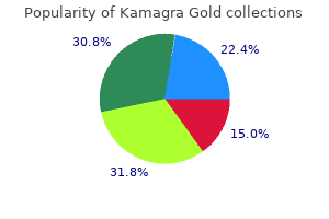
Kamagra Gold dosages: 100 mg
Kamagra Gold packs: 30 pills, 60 pills, 90 pills, 120 pills, 180 pills

Kamagra gold 100 mg amex
The crystals were partly dissolved when the paraffin block was softened by 10% ammonia resolution erectile dysfunction 55 years old buy generic kamagra gold 100 mg on-line. By energy-dispersive X-ray spectroscopy erectile dysfunction treatment natural kamagra gold 100 mg cheap with amex, the crystals had been proven to be natural in nature, containing carbon but neither calcium nor sodium, and immunohistochemical results argued towards keratin tonofilament origin. There are small and enormous, tender, infected nodules, cysts, and discharging sinuses that finally heal, leaving disfiguring scars. Doxycycline or oral trimethoprim� sulfamethoxazole is used for secondary infections. It is often confined to the beard area of the face and neck,493 but not often the scalp,494 pubic space,495 and legs496 may be concerned. Hair shafts in black individuals have a tendency to form tight coils and, following shaving, the sharp ends may pierce the skin adjoining to the orifice of the follicles. Small overseas physique granulomas and a blended inflammatory cell infiltrate are current in older lesions. In some instances, epithelium grows down from the surface to encase both the hair and the inflammatory response, assisting of their eventual transepithelial elimination. Dermoscopy is comparatively distinctive, exhibiting a gray-blue, thick curved line and adjacent pink traces (probably reactive vessels) upon a structureless pattern. A variable inflammatory response is current in the dermis in this region, and sometimes a granulomatous perifolliculitis develops. A deep scarring, or profunda, kind has been described in which perforation occurs at a number of different ranges of the follicle, with a extra extensive granulomatous infiltrate and destruction of the follicular epithelium and sebaceous gland. Most cases have been associated with the ingestion of medication, particularly antibiotics, though in others an enterovirus infection has been incriminated. Histopathology198,508 There is a dilated follicular infundibulum full of keratinous and mobile particles. The walls of the hair follicles are enlarged and irregularly deformed with their epithelial define blurred by a lymphocytic infiltrate. Of the 15 instances reported in a single collection, 10 were composed predominantly of B cells and the remainder predominantly of T cells. Other cell sorts in addition to lymphocytes included plasma cells and epithelioid macrophages. Three of 30 tested instances were constructive for herpes simplex virus-1, a condition identified to be associated with atypical lymphoid infiltrates. Rearrangements of T-cell receptor genes (19 cases) and immunoglobulin heavy-chain genes (3 cases) had been identified; there have been no variations between clonal and nonclonal cases by means of medical, histopathologic, or immunohistochemical options. Sometimes, nonetheless, these abnormalities occur sporadically or intermittently,543 involving only isolated hairs as an incidental phenomenon. This is particularly likely to occur in those who topic their hair to physical or chemical trauma. Graying seems to be a consequence of an general and specific depletion of melanocytes in the hair bulbs and the outer root sheath. In that case, a 7-cm-long submerged hair extended as a blue line just under the skin surface. Other examples of this phenomenon, resulting from implantation of exogenous hair shafts, have been reported. One case involved the glans penis,541 and one other involved the only of the foot of a child. Scalp hair is most frequently concerned, though the genital area can also be affected. A case diagnosed by trichoscopy (a type of dermoscopy) was shown to be associated with nail dystrophy. In black people, trauma-related instances of trichorrhexis nodosa usually affect the proximal part of the hair shaft, whereas in white and Oriental races, the distal half is extra often affected. Histopathology There is a cup-like expansion of the proximal part of the hair shaft that surrounds the club-shaped distal section within the method of a balland-socket joint. Paraffin-blocked hairs have a triangular or kidney-shaped look on transverse part. A common change, which is usually classified as a discrete abnormality, is longitudinal grooving of the hair. Included in this group are the following entities: � Pili canaliculati et trianguli � Pili bifurcati � Pili multigemini � Trichostasis spinulosa � Pili annulati � Monilethrix � Tapered hairs � Bubble hair � Trichomegaly. It differs from tufted-hair folliculitis, in which distinctive tufts of multiple hairs emerge from a single follicular orifice into which several full follicles open (see p. There is retention of telogen hairs throughout the follicles, though the reason for that is unknown. Straight hair nevus, a localized disorder in which the concerned hairs are brief and straight � in distinction to the same old woolly hair of black folks, in whom this situation has been described � has been thought to be a localized form of uncombable hair. There is only one hair matrix and papilla on the base of the follicle,530 in contrast to pili multigemini. Transmission electron microscopy has proven an irregular cortex with areas of homogeneous nonfibrillar materials and a deviated axis of some microfibrils. It may be an autosomal dominant dysfunction producing hypotrichosis of the whole scalp or simply the occipital area, an acquired situation related to numerous hair fragility disorders, and an iatrogenic condition. The Pohl�Pinkus mark is an isolated narrowing of the hair shaft that coincides with a surgery or some other traumatic episode. It may be associated with extra generalized hypertrichosis or with involvement of the eyebrows as properly. An wonderful review of eyelash trichomegaly and its disease associations has been written by Paul and colleagues. The condition of pili torti is the most important member of this group, whereas trichonodosis and circle hairs are of little consequence. Pili torti could occur alone or in association with different syndromes, notably of ectodermal kind. An acquired form of pili torti has been described on account of trauma, related to cicatricial alopecia, following using synthetic retinoids (isotretinoin),659 and on the stomach and thighs of hirsute women and men. Curvatures and twisting have been recorded within the hair follicles that produce pili torti. There is a diffuse partial form, during which shorter, finer, curly hair is interspersed with normal hair,664 and a well- Histopathology There is usually some flattening of the hair shafts with partial twisting at irregular intervals. In reality, the absence of curly hair on this syndrome is related to a milder phenotype, with higher motor operate. African hair length is lower than that of white folks, probably because of the hair loss related to the combing of knotted hair. Various hair shaft abnormalities of a secondary nature may be current in and adjoining to the knots. It has many causes other than trichonodosis, together with the following: hair density and coiling; chemical and physical treatments to the hair, including using conditioners;691 frequent combing;692 and neglect.

Order kamagra gold 100 mg fast delivery
Amoxicillin-induced exanthema in young adults with infectious mononucleosis: Demonstration of drug-specific lymphocyte reactivity erectile dysfunction history 100 mg kamagra gold purchase amex. Slow acetylator genotypes as a possible threat factor for infectious mononucleosis-like syndrome induced by salazosulfapyridine erectile dysfunction drug related kamagra gold 100 mg generic without prescription. Adverse cutaneous reactions to trimethoprim sulfamethoxazole in sufferers with the acquired immunodeficiency syndrome and Pneumocystis carinii pneumonia. Which intercurrent infections are related to maculopapular cutaneous drug reactions Codeine brought on pruritic scarlatiniform exanthemata: Patch test negative but optimistic to oral provocation check. Morbilliform eruptions attributable to penicillin: A examine by electron microscopy and immunologic tests. Histopathologic options of exanthematous drug eruptions of the macular and popular kind. Iododerma after computed tomographic scan with intravenous radiopaque distinction media. Potassium iodide in dermatology: A nineteenth century drug for the 21st century � Uses, pharmacology, opposed results, and contraindications. Heydenreich G, Olholm Larsen P Iododerma after excessive dose urography in an oliguric. Dapsone hypersensitivity syndrome in a affected person with cutaneous lupus erythematosus. Allopurinol hypersensitivity syndrome: Hypersensitivity to oxypurinol but not allopurinol. Sequential evaluation of an antidrug antibody response in a patient with a systemic delayed-onset sulphonamide hypersensitivity syndrome reaction. A close to fatal case of the dapsone hypersensitivity syndrome in a patient with urticarial vasculitis. Clinicopathological features and prognosis of drug rash with eosinophilia and systemic signs: A research of 30 instances in Taiwan. Anticonvulsant hypersensitivity syndrome related to Bellamine S, a therapy for menopausal symptoms. Severe sialadenitis: A new complication of drug reaction with eosinophilia and systemic symptoms. Clopidogrel-induced hypersensitivity syndrome related to febrile pancytopenia. Drug-induced hypersensitivity syndrome related to Epstein�Barr virus an infection. Anticonvulsant hypersensitivity syndrome related to reactivation of cytomegalovirus. Carbamazepine-induced hypersensitivity syndrome associated with transient hypogammaglobulinaemia and reactivation of human herpesvirus 6 an infection demonstrated by real-time quantitative polymerase chain reaction. Association of human herpesvirus 6 reactivation with the flaring and severity of drug-induced hypersensitivity syndrome. Association between anticonvulsant hypersensitivity syndrome and human herpesvirus 6 reactivation and hypogammoglobulinemia. A case of drug-induced hypersensitivity syndrome showing transient immunosuppression earlier than viral reactivation during remedy for pemphigus foliaceus. Increased levels of interleukin 5 are associated with the era of eosinophilia in drug-induced hypersensitivity syndrome. Fulminant type 1 diabetes mellitus caused by drug hypersensitivity syndrome with human herpesvirus 6 an infection. Syndrome of inappropriate secretion of antidiuretic hormone related to limbic encephalitis in a affected person with drug-induced hypersensitivity syndrome. Drug-reaction eosinophilia and systemic signs and drug-induced hypersensitivity syndrome. Sodium valproate-induced cutaneous pseudolymphoma followed by recurrence with carbamazepine. Cutaneous pseudolymphoma due to benidipine hydrochloride with massive infiltration of eosinophils. Cutaneous pseudolymphoma induced by adalimumab and reproduced by infliximab in a affected person with arthropathic psoriasis. Drug-induced immune dysregulation as a reason for atypical cutaneous lymphoid infiltrates: A hypothesis. Dermatological antagonistic effects with the antimalarial drug mefloquine: A review of 74 revealed case reviews. Cutaneous opposed reactions to clindamycin: Results of skin checks and oral publicity. Serious antagonistic reactions induced by minocycline: Report of 13 patients and evaluate of the literature. Fever, lymphadenopathy, eosinophilia, lymphocytosis, hepatitis, and dermatitis: A extreme adverse response to minocycline. Efficacy and security of desensitization with sulfamethoxazole and trimethoprim in forty eight previously hypersensitive sufferers infected with human immunodeficiency virus. Macrolides, fluoroquinolones, rifamycins, tetracyclines, trimethoprim� sulfamethoxazole, and clindamycin. Adverse drug reactions of the new antifungal brokers � Terbinafine, fluconazole, and itraconazole. Safety of oral terbinafine: Results of a postmarketing, surveillance study in 25884 patients. Drug interactions with itraconazole, fluconazole, and terbinafine and their administration. Drug eruption secondary to aciclovir with recall phenomenon in a dermatome beforehand affected by herpes zoster. An expanded profile of cutaneous reactions to nonsteroidal anti-inflammatory medicine. Fixed drug eruption associated to ibuprofen presenting as big bullae on the posterior thigh. Acute generalized exanthematous pustulosis-like, folliculitic drug response pattern caused by celecoxib. Lamotrigine and phenobarbitone-associated hypersensitivity syndrome: Resolution with out corticosteroids. Interferon- manufacturing within the peripheral lymphocytes of a patient with carbamazepine hypersensitivity syndrome. Generalized pustulation as a manifestation of the anticonvulsant hypersensitivity syndrome. Generalized pustulosis and severe tubulointerstitial nephropathy as manifestations of cabamazepine hypersensitivity syndrome.
Order kamagra gold 100 mg with visa
Basal cell carcinomas of the inside canthus: Incidence of incomplete excision according to zocor impotence 100 mg kamagra gold purchase mastercard topographical localization of tumours erectile dysfunction drugs non prescription discount kamagra gold 100 mg with mastercard. Recurrent morphoeic basal cell carcinoma at the lateral canthus with orbitocranial invasion. Correlation of histologic subtypes of major basal cell carcinoma and variety of Mohs levels required to achieve a tumor-free plane. Histologic evolution of recurrent basal cell carcinoma and treatment implications. Basal cell carcinoma: Tumor clustering is associated with elevated accrual in high risk subgroups. Basal cell carcinoma recurring after radiotherapy: A distinctive, tough remedy subclass of recurrent basal cell carcinoma. Syndecan-1 expression is decreased with growing aggressiveness of basal cell carcinoma. Altered expression of the hemidesmosome-anchoring filament complicated proteins in basal cell carcinoma: Possible position within the origin of peritumoral lacunae. Overexpression of protein kinase C- and - isozymes by stromal dendritic cells in basal and squamous cell carcinoma. Correlation of the expression of human telomerase subunits with telomerase activity in normal pores and skin and pores and skin tumors. The prognostic significance of tumor angiogenesis in nonaggressive and aggressive basal cell carcinoma of the human pores and skin. Differences in tumor microvessel density between squamous cell carcinomas and basal cell carcinomas could relate to their different biologic behavior. Metastatic basal cell carcinoma: Report of five circumstances and evaluation of one hundred seventy cases within the literature. Basal cell carcinoma of the scalp resulting in spine metastasis in a black patient. Metastatic basal cell carcinoma with squamous differentiation: Report of a case with response of cutaneous metastases to electrochemotherapy. Metastatic basal cell carcinoma presenting with unilateral upper extremity edema and lymphatic spread. Metastatic basal cell carcinoma: Four case reports, review of literature, and immunohistochemical evaluation. Metastatic basal cell carcinoma: Report of twelve circumstances with a review of the literature. Giant basal cell carcinoma with metastasis and secondary amyloidosis: Report of case. Differentiation of basal cell carcinoma from trichoepithelioma by lectin histochemistry. The fee and pattern of bcl-2 and cytokeratin 15 expression in trichoepithelioma and nodular basal cell carcinoma: A comparative examine. Stromelysin-3: A potent marker for histopathologic differentiation between desmoplastic trichoepithelioma and morphealike basal cell carcinoma. Fibroblast-activation protein: A single marker that confidently differentiates morpheaform/infiltrative basal cell carcinoma from desmoplastic trichoepithelioma. Diagnostic utility of cytokeratin 17 immunostaining in morpheaform basal cell carcinoma and for facilitating the detection of tumor cells at the surgical margins. Immunohistologic differential prognosis of basal cell carcinoma, squamous cell carcinoma, and trichoepithelioma in small cutaneous biopsy specimens. Familial basaloid follicular hamartoma: Lesional characterization and evaluation of the literature. Primary adenoid cystic carcinoma of the pores and skin: A medical, histological, and immunocytochemical comparability with adenoid cystic carcinoma of salivary glands and adenoid basal cell carcinoma. Primary cutaneous cribriform apocrine carcinoma: A clinicopathologic and immunohistochemical examine of 26 circumstances of an under-recognized cutaneous adnexal neoplasm. Dermal adjustments in superficial basal cell carcinoma, melanoma in situ and actinic keratosis and their implications. Use of a murine monoclonal antibody which binds to malignant keratinocytes to detect tumor cells in microscopically managed surgical procedure. Involucrin in squamous and basal cell carcinomas, of the pores and skin: An immunohistochemical examine. Naevoid basal cell carcinoma syndrome with a palmar epidermoid cyst, milia and maxillary cysts. Atlanto-occipital ligament calcification: A novel sign in nevoid basal cell carcinoma syndrome. Nevoid basal cell carcinoma syndrome: Bilateral ovarian fibromas in a three 12 -year-old girl. Nevoid basal cell carcinoma syndrome associated with unilateral renal agenesis: Acceleration of basal cell carcinomas following radiotherapy. Basal cell nevus syndrome concurrent with adenoid cystic carcinoma of salivary gland. Patched homologue 1 mutations in four Japanese families with basal cell nevus syndrome. Fine mapping of the locus for nevoid basal cell carcinoma syndrome on chromosome 9q. Basal cell carcinoma occurring in a quantity of familial trichoepithelioma: Detection of lack of heterozygosity in chromosome 9q. Nonsense-associated altered splicing of the, Patched gene fails to suppress carcinogenesis in Gorlin syndrome. Nevoid basal cell carcinoma syndrome: A 15-year follow-up of instances in Ottawa and the Ottawa Valley. Nevoid basal cell carcinoma syndrome: Multiple basal cell carcinomas of the palm after radiation remedy. Multiple polypoid basal cell carcinomas on the perineum of a affected person with basal cell nevus syndrome. Sun publicity and basal cell carcinomas within the nevoid basal cell carcinoma syndrome. The nevoid basal cell carcinoma syndrome: Sensitivity to ultraviolet and X-ray irradiation. Nevoid basal cell carcinoma syndrome: Profile of genetic and environmental components in oncogenesis. Unilateral linear basal cell nevus related to diffuse osteoma cutis, unilateral anodontia, and irregular bone mineralization. Congenital linear unilateral basal cell nevus: A case report with patched gene molecular research. Linear unilateral hamartomatous basal cell naevus with glandular and follicular differentiation. Multiple familial basal cell carcinomas together with, a case of segmental manifestation. Tissue and tumor mosaicism of the myotonin protein kinase gene trinucleotide repeat in a affected person with multiple basal cell carcinomas associated with myotonic dystrophy. A novel phenotype with options of basal, cell nevus syndrome and basaloid follicular hamartoma syndrome.

Generic kamagra gold 100 mg amex
Fatal end result of generalized morphoea with bronchiolitis obliterans organizing pneumonia erectile dysfunction diabetes qof kamagra gold 100 mg amex. Lymphoproliferative responses to Borrelia burgdorferi in circumscribed scleroderma erectile dysfunction drugs and heart disease buy 100 mg kamagra gold with amex. Spirochetal varieties within the dermal lesions of morphea and lichen sclerosus et atrophicus. Histological proof of spirochetal origin of morphea and lichen sclerosus et atrophicans. A morphealike pores and skin situation attributable to Borrelia burgdorferi in an immunocompromised patient. Do some sufferers with morphea and lichen sclerosus et atrophicans have a Borrelia an infection Antibodies to the Borrelia burgdorferi flagellum in sufferers with scleroderma, granuloma annulare and porphyria cutanea tarda. No evidence for a relation between Borrelia burgdorferi infection and old lesions of localized scleroderma (morphea). Morphoea is neither associated with options of Borrelia burgdorferi infection, nor is this agent detectable in lesional skin by polymerase chain response. IgG antibody reactivity to Borrelia burgdorferi sensu stricto antigens in sufferers with morphea in Colombia. Immunohistochemical analysis of remodeling growth factor three expression in solitary morphoea profunda with histological membranocystic adjustments. Serum concentration of procollagen sort 1 carboxyterminal propeptide in localized scleroderma. Expression of insulin-like development factor-I in lesional and non-lesional skin of patients with morphoea. Clinical characteristics related to antihistone antibodies in sufferers with localized scleroderma. Rheumatoid factor isotypes and anti-agalactosyl IgG antibodies in systemic sclerosis. Increased levels of circulating intercellular adhesion molecule-1 in sufferers with localized scleroderma. Differential expression of decorin in localized scleroderma following ultraviolet-A1 irradiation. Epithelial�mesenchymal transition of the eccrine glands is concerned in pores and skin fibrosis in morphea. Localized scleroderma in breast most cancers patients treated with supervoltage exterior beam radiation: Radiation port scleroderma. Postirradiation morphea of the breast: Presentation of two cases and evaluation of the literature. Drug-induced morphea: Report of a case induced by balicatib and evaluate of the literature. Morphea associated with the use of adalimumab: A case report and evaluation of the literature. Morphea-like pores and skin reactions in sufferers handled with the cathepsin K inhibitor balicatib. Treatment of plaque-type localized scleroderma with retinoic acid and ultraviolet A plus the photosensitizer psoralen: A case series. Combined remedy with calcipotriol ointment and low-dose ultraviolet A1 phototherapy in childhood morphea. First case sequence on using calcipotriol� betamethasone dipropionate for morphea. Pulsed high-dose corticosteroids mixed with low-dose methotrexate in severe localized scleroderma. Topical imiquimod 5% cream for pediatric plaque morphea: A potential, multiple-baseline, open-label pilot research. Abatacept is a promising therapy for sufferers with disseminated morphea profunda: Presentation of two circumstances. Clinical and serologic expression of localized scleroderma: Case report and evaluate of the literature. Linear scleroderma: Report of a case presenting as persistent unilateral eyelid edema. Scleroderma en coup de sabre with central nervous system and ophthalmologic involvement: Treatment of ocular symptoms with interferon gamma. Brain cavernomas related to en coup de sabre linear scleroderma: Two case stories. Progressive hemifacial atrophy with scleroderma and ipsilateral limb losing (Parry�Romberg syndrome). Clinical and serological traits of progressive facial hemiatrophy: A case sequence of 12 sufferers. Long-lasting follow-up favours an in depth relationship between progressive facial hemiatrophy and scleroderma en coup de sabre. Linear scleroderma related to hypertrichosis within the absence of melorheostosis. Immunohistochemical characterization of the mobile infiltrate in localized scleroderma. Distinctive histopathologic findings in linear morphea (en coup de sabre) alopecia. Ossification in linear morphoea with hemifacial atrophy � Treatment by surgical excision. Bullous morphea: Clinical, pathologic, and immunopathologic evaluation of 13 circumstances. Morphea limited to the superficial reticular dermis: An underrecognized histologic phenomenon. A clinical, histological, and immunohistochemical comparability of acrodermatitis chronica atrophicans and morphea. Analysis of dendritic cell populations using a revised histological staging of morphoea. Self-involuting atrophoderma of the lateralupper arm: A new benign variant of morphea Diagnostic usefulness of dermatoscopy in differentiating lichen sclerosus et atrophicus from morphea. Clinical and dermoscopic findings of a affected person with co-existing lichen planus, lichen sclerosus and morphea. Nodular scleroderma: Focally increased tenascin expression differing from that in the surrounding scleroderma skin. Cutaneous sclerosis associated with left ventricular noncompaction, myopathy, polyneuropathy and pancytopenia with eosinophilia. Scleroderma-like pores and skin sclerosis induced by docetaxel chemotherapy for hormone refractory prostate most cancers: A case report.

100 mg kamagra gold quality
They have a attribute histological appearance with dense collagen bundles erectile dysfunction diagnosis code generic 100 mg kamagra gold mastercard, usually in a storiform association erectile dysfunction due diabetes buy kamagra gold 100 mg amex. One such instance is the dermal nodule found within the sacrococcygeal region of bicycle riders. There is superficial telangiectasia and/or lymphangiectasia with delicate epidermal atrophy. There is commonly a mild deep perivascular infiltrate of lymphocytes, however this was not current in two instances seen by the writer. Connective tissue nevi and hypertrophic scars and keloids have been arbitrarily included on this section; they may be considered tumor-like proliferations of fibrous tissue, different examples of that are discussed in Chapter 34. Sometimes there are alterations in multiple component of connective tissue, and these lesions might simply be categorized as connective tissue nevi. Histopathology the epidermis is usually normal, though an overlying epidermal nevus has been reported. They usually current as asymptomatic, firm, flesh-colored plaques and Shagreenpatch the shagreen patch is a distinct scientific variant of collagenoma, found exclusively in these with tuberous sclerosis. There is a circumscribed area of thickened collagen bundles, involving the papillary and mid dermis. A review summarizes the roles of varied growth components, cytokines, extracellular matrix proteins, and proteolytic enzymes involved within the formation of hypertrophic scars. Early lesions are erythematous, whereas older lesions are normally pale, although sometimes they might be pigmented. Attempted surgical excision of a keloid ends in regrowth of a bigger lesion unless some concurrent efficient therapeutic measures, corresponding to radiation remedy or the use of cultured epithelial autografts, are adopted. A study of 14 pedigrees with familial keloids showed an autosomal dominant mode of inheritance with incomplete clinical penetrance and variable expression. A case of segmental tuberous sclerosis has been reported by which there have been unilateral angiofibromas and periungual fibromas in addition to a shagreen patch. The overlying dermis is usually flat, although typically it has the sample of acanthosis nigricans. Fibroblasts isolated from keloids typically synthesize regular quantities of collagen,825 although proline hydroxylase activity, a marker of collagen synthesis, is higher in keloids than in hypertrophic scars or normal wounds. It could possibly be the effector mechanism for the regression that outcomes from mechanical compression. Topical therapies used embody pressure therapy,861 silicone gel sheeting and ointment,862 polyurethane dressing, onion extract, imiquimod 5% cream, and vitamins A and E. Asiaticoside, a by-product of the plant Centella asiatica (Indian pennywort), has been used for the therapy of hypertrophic scar for many years as an ingredient of Gotu Kola, a traditional herbal drugs. Fibroblasts are increased and are discovered alongside the collagen bundles with an orientation much like the accompanying collagen. Keloids have reduced vascularity in contrast with hypertrophic scars and regular healing wounds. The overlying epidermis may be skinny, and beneath it there are sometimes some telangiectatic vessels. A sparse, persistent inflammatory cell infiltrate might encompass these peripheral vessels. Capillaries are typically oriented perpendicular to the pores and skin surface, and these could additionally be surrounded by a sparse inflammatory cell infiltrate within the scar. Dystrophic calcification and bone may occasionally develop, significantly in stomach scars. The dermis could also be thinned, suggesting that this condition would be better considered with the atrophic collagenoses. The elastin and fibrillin fibers in the deep dermis show realignment parallel to the pores and skin surface. They have been Differential analysis Keloids are sufficiently distinctive that microscopic prognosis is seldom a problem. However, the attribute broad, homogeneous, haphazardly distributed, eosinophilic collagen bundles of keloid can often be noticed focally in lesions that in any other case have the general contours of ordinary or hypertrophic scars. Other fibrotic or sclerosing conditions, including morphea and dermatofibroma, can also present keloidal areas. However, these conditions can typically be excluded by scientific knowledge or by chosen microscopic features. Attention has been drawn just lately to the presence of S100-positive cells, together with spindle cells with gentle atypia, in cutaneous scars. They are significantly more common on the thighs of girls and on the knees of boys. Another study instructed that the early levels consist of mast cell degranulation, followed by an inflow of activated macrophages that envelop fragmented elastic fibers. Reflectance confocal microscopy exhibits parallel collagen bundles working perpendicular to the lengthy axis of the lesions and parallel fantastic papillary dermal collagen bundles with oval-shaped dermal papillae within lesions, in contrast to rounded dermal papillae in surrounding regular pores and skin. Thinner collagen bundles had been seen in striae rubra, in contrast to these in striae alba. The levels of cellularity range from a density suggestive of fibromatosis to extra sparsely mobile lesions. Smooth muscle actin staining, indicative of myofibroblasts, is only variably positive amongst spindle cells. There is considerable involvement of the subcutis, and this entity is subsequently considered in additional detail with the panniculitides (see Chapter 17, p. Reported circumstances have all been in elderly males with an outside occupation or interest. They differ from elastotic nodules of the ear, which have severe actinic elastosis, not cartilaginous metaplasia with a spur of fibrous tissue as seen in weathering nodules. The collagen merges almost imperceptibly with the decrease dermis and the ligamentum nuchae. Furthermore, there are only a few fibroblasts, distinguishing the lesion from a fibromatosis. However, there has been a report of a desmoid fibromatosis having areas resembling collagenosis nuchae. Technical artifacts within the preparation of histological sections can even affect dermal thickness. The following circumstances, discussed here, could be associated with a decrease in dermal thickness: 370 Section4 � TheDermisandSubcutis � Aplasia cutis congenita � Focal dermal hypoplasia � Focal facial dermal dysplasia � Pseudoainhum constricting bands � Keratosis pilaris atrophicans � Corticosteroid atrophy � Linear atrophoderma of Moulin � Acrodermatitis chronica atrophicans � Restrictive dermopathy. The distinctive case with membranocystic degeneration of collagen fibers, associated with some deposition of fat, and with the clinical look of xanthomatosis, defies classification. This syndrome is distinct from focal dermal hypoplasia, but it could possibly be included with focal facial dermal dysplasia (see later).

Purchase kamagra gold 100 mg otc
Perifolliculitis capitis abscedens et erectile dysfunction jack3d discount 100 mg kamagra gold with visa, suffodiens efficiently managed with infliximab erectile dysfunction treatment milwaukee kamagra gold 100 mg order mastercard. Acne conglobata of the buttocks aggravated by mechanical and environmental components. Acne conglobata and a generalized lichen spinulosus-like eruption in a man seropositive for human immunodeficiency virus. Single nucleotide polymorphisms of toll-like receptor-4 protect in opposition to zits conglobata. Cutaneous side-effects in most cancers patients, treated with the antiepidermal development issue receptor antibody C225. Necrotizing infundibular crystalline folliculitis manifesting as a perforating mucinosis: A case report. Epiluminescence dermatoscopy enhanced affected person compliance and achieved treatment success in pseudofolliculitis barbae. Pityrosporum folliculitis during being pregnant: A attainable reason for pruritic folliculitis of pregnancy. Perforating folliculitis with jaundice in an Indian male: A uncommon case with sclerosing cholangitis. Perforating folliculitis associated with tumour necrosis factor- inhibitors administered for rheumatoid arthritis. Perforating folliculitis in a patient treated with nilotinib: A additional evidence of c-kit involvement. Sterile neutrophilic folliculitis with perifollicular vasculopathy: A distinctive cutaneous reaction pattern reflecting systemic illness. Pseudolymphomatous folliculitis: A clinicopathologic examine of 15 circumstances of cutaneous pseudolymphoma with follicular invasion. A review of fifty five instances of cutaneous lymphoid hyperplasia: Reassessment of the histopathologic findings leading to reclassification of four lesions as cutaneous marginal zone lymphoma and 19 as pseudolymphomatous folliculitis. Hyperplasia of hair follicles and different adnexal structures in cutaneous lymphoproliferative problems: A examine of fifty three instances, together with so-called pseudolymphomatous folliculitis and overt lymphomas. Light microscopic examination of scalp hair samples as an assist within the prognosis of paediatric disorders: Retrospective evaluation of greater than 300 cases from a single centre. Human hair greying is linked to a selected depletion of hair follicle melanocytes affecting both the bulb and the outer root sheath. Trichotemnomania related to trichotillomania: A case report with emphasis on the diagnostic value of dermoscopy. Trichothiodystrophy: Review of sulfur-deficient brittle hair syndromes and affiliation with the ectodermal dysplasias. Intractable diarrhea of infancy with facial dysmorphism, trichorrhexis nodosa, and cirrhosis. Characterization of tiger tail banding and hair shaft abnormalities in trichothiodystrophy. Trichothiodystrophy: Sulfur-deficient brittle hair as a marker for a neuroectodermal symptom advanced. Trichothiodystrophy: Clinical spectrum, central nervous system imaging, and biochemical characterization of two siblings. Trichothiodystrophy associated with photosensitivity, gonadal failure, and putting osteosclerosis. Cheveux incoiffables � Diagnostic, clinical and, hair microscopic findings, and pathogenic research. Uncombable hair syndrome: Observations on response to biotin and prevalence in siblings with ectodermal dysplasia. Pili canaliculi: Clinical and microscopic investigation of the primary Brazilian family. Quantitative assessment of scanning electron microscope defects in uncombable-hair syndrome. Straight hair related to interferon-alfa plus, ribavirin in hepatitis C infection. Pili trianguli et canaliculi is a defect of inner root sheath keratinization: Ultrastructural observations of anomalous tonofilament group in a case. Trichostasis spinulosa: Possible association with extended topical software of clobetasol proprionate 0. Study of nanomechanical properties of, human hair shaft in a case of pili annulati by atomic drive microscopy. Concomitant manifestation of pili annulati and alopecia areata: Coincidental somewhat than true association. Pili annulati coincident with alopecia areata, autoimmune thyroid illness and first IgA deficiency: Case report and considerations on the literature. Newly described weathering sample in, pili annulati hair shafts: A scanning electron microscopic study. Pseudopili annulati in a dark-haired individual: A gentle and electron microscopic examine. Update on detection, morphology and fragility, in pili annulati in three kindreds. Alterations within the basement membrane zone in pili annulati hair follicles as demonstrated by electron microscopy and immunohistochemistry. Linkage of monilethrix to the trichocyte, and epithelial keratin gene cluster on 12q11�q13. A mutational hotspot within the 2B area of, human hair primary keratin 6 (hHb6) in monilethrix sufferers. Identification of novel mutations in primary hair, keratins hHb1 and hHb6 in monilethrix: Implications for protein structure and clinical phenotype. Severe monilethrix related to intractable scalp pruritus, posterior subcapsular cataract, brachiocephaly, and distinct facial features: A new variant of monilethrix syndrome Bubble hair: Case attributable to an overheating, hair dryer and reproducibility in normal hair with warmth. Bubble hair: A cosmetic abnormality attributable to temporary, focal heating of damp hair fibres. Bubble hair and different acquired hair shaft anomalies as a result of hot ironing on moist hair. Facial hypertrichosis and trichomegaly creating in sufferers handled with the epidermal progress issue receptor inhibitor erlotinib. Eyelash trichomegaly: Review of congenital, acquired, and drug-associated etiologies for elongation of the eyelashes. Three members of a household with pili torti and sensorineural listening to loss: the Bjornstad syndrome. Cutaneous findings in a new syndrome of autosomal recessive ectodermal dysplasia with corkscrew hairs. Autosomal-dominant woolly hair ensuing from disruption of keratin 74 (krt74), a potential determinant of human hair texture. Woolly-hair nevus: Report of a case associated with a verrucous epidermal nevus in the same area. Systematized, unilateral, velvety hyperpigmentation and homolateral patches of curled hairs.
Kamagra gold 100 mg generic with visa
Another problematic scenario is presented by the ocular melanoma with blue nevus-like metastases to distant cutaneous sites erectile dysfunction causes n treatment purchase 100 mg kamagra gold mastercard. Histopathology In each dermal melanocyte hamartoma and phakomatosis pigmentovascularis erectile dysfunction medication levitra buy kamagra gold 100 mg lowest price, there are moderate numbers of dendritic melanocytes scattered throughout the higher and mid dermis, similar to the macular areas of the nevus of Ota. The diagnosis is made by finding cytological features of malignancy, similar to nuclear pleomorphism and atypical mitoses, mixed with subcutaneous invasion and sometimes some necrosis. The presence of atypical mitoses seems to be a more specific function of malignancy than necrosis. Cytological atypia and frequent mitoses could also be seen, however necrosis and atypical mitoses are normally absent, distinguishing such cases from malignant blue nevus. Both are significantly totally different from that seen in benign nevomelanocytic lesions. A fifth type (type V) with cutis marmorata and dermal melanocytosis was added in 2003. A rare acquired variant of dermal melanocytosis (acquired dermal melanocytosis) has been reported. Follow-up information indicated that every one patients have been alive with out illness after a period of 35�92 months (mean, fifty four months). The lesions, which resemble blue nevi or congenital nevi clinically, average 3�7 cm in diameter. The scalp is a standard website of involvement;999�1001 one case offered as cutis verticis gyrata. Histopathology Cutaneous neurocristic hamartoma most resembles a congenital nevus with neuroid options. There is a variable admixture of nevomelanocytes, Schwann cells, and dendritic blue nevus cells. Not all nevi with these scientific characteristics have the histological features of dysplastic (atypical) nevi (see later). In sufferers with the dysplastic (atypical) nevus syndrome, the number of nevi is large � up to 80 or extra. In childhood, nevi normally seem regular; the abnormal lesions appear in adolescence and grownup life. The dysplastic (atypical) nevus trait has a more sophisticated inheritance, occurring more commonly than is common for dominant inheritance. If such a lesion warrants a biopsy, it ought to be an entire excisional biopsy (which can include shave or punch) to permit full sampling of the lesion. Random cytological atypia refers to the presence of occasional cells with enlarged hyperchromatic nuclei, generally with prominent nucleoli. The nuclei equal the nucleus of the overlying keratinocytes in dimension or are bigger. There can also be a patchy superficial lymphocytic infiltrate1040 and, generally, new vessel formation. Neutrophils have been described around some of the epidermal and dermal melanocytic nests in a couple of circumstances. In different phrases, dysplastic (atypical) nevi are normally compound nevi with peripheral lentiginous and junctional activity and random cytological atypia in the epidermal element. Toussaint and Kamino examined a big sequence of dysplastic nevi and located that the dermal element of some instances may show features of different varieties of nevi, similar to a congenital nevus, Spitz nevus, blue nevus, halo nevus, or dermal neuronevus. The malignant lesion that may come up in a cutaneous neurocristic hamartoma is discussed on web page 863. Before age forty years, the incidence is greater in women, with most melanomas occurring on the lower extremities and exhibiting the superficial spreading histology. These irregular melanosomes are transferred to keratinocytes before being completely melanized, they usually reveal marked degradation. Some still think about this an important precursor of melanoma and melanoma in situ in the elderly. Similar lesions have been categorized in the past (and presumably nonetheless are in most centers) as dysplastic nevus, atypical junctional nevus, melanoma in situ (early or evolving), and premalignant melanosis. Primary mucosal melanomas are commonest in the nasal cavity, sinuses, oropharynx, anorectum, vulva (including clitoris and labia), and vagina. Primary melanomas have additionally been reported within the prostate,1220,1221 salivary glands,1222,1223 kidney,1224 thyroid,1225 thymus,1226 pancreas,1227 ovary,1228,1229 and adrenal glands,1230,1231 with many cases doubtless arising in extensions of the mucosal network. It is unclear what quantity of of these reported cases are true main melanomas and not metastases with an occult main. In general, extracutaneous melanomas have a much worse prognosis than their cutaneous counterparts. Riskfactors Genetic and environmental elements arguably play a job in the epidemiology of any illness, including melanoma. William Norris, a common practitioner in the United Kingdom, recognized this fact when he described a family with numerous moles and melanoma and subsequently described eight melanoma patients, some with gentle hair, pale complexion, and attainable industrial pollution exposure. Many 868 Section7 � Tumors studies have examined the sun-exposure habits of patients with melanoma. Studies have demonstrated a positive correlation between sunscreen use and a decrease in melanoma incidence, but other studies have shown no correlation or even a adverse correlation. Sun avoidance and sun safety remain the mainstays of melanoma prevention for persons at high risk because no clear selection for effective melanoma chemoprevention has emerged. The risk significantly increases with younger age of first publicity and repetitive use. Reported associations include publicity to polyvinyl chloride,1285,1286 pesticides,1287�1289 chemical solvents,1290,1291 arsenic-polluted water,1292,1293 and redox-active metals (occupational exposures and/or joint replacements),1294 amongst different exposures. Proneness to develop nevi correlates not only with sun exposure, including blistering episodes, but in addition with skin complexion, hair colour, and tanning capacity. A study from Boston printed in 2003 estimated that the annual transformation rate of any single nevus right into a melanoma ranges from zero. Some of those could symbolize the chance incidence of two situations, whereas others are extra agency as new genetic data are rising. Family history is variably outlined, nevertheless it usually features a single first-degree member of the family or multiple more distant family members affected on the identical side of the family. In a person with a constructive household historical past, the risk varies greatly relying on the circumstances. A constructive household history equates to roughly a twofold threat in creating melanoma. All of these reasons introduce potential factors of intervention by elevated surveillance. Melanoma susceptibility genes play roles in cell cycling, melanocyte development, and melanin biosynthesis. They can be subclassified into high-, intermediate-, and low-risk genes, and excellent evaluations are available on this topic.
Kamagra gold 100 mg purchase online
The efficacy of biologic brokers erectile dysfunction and diabetes 100 mg kamagra gold cheap free shipping, notably tumor necrosis factor- blockers xarelto impotence buy discount kamagra gold 100 mg on line, has not been decided with certainty, partly due to the absence of randomized managed trials and completely different treatment end factors used within the numerous present publications. The inflammatory cell infiltrate initially contains many neutrophils, however there are progressively extra lymphocytes, plasma cells, and histiocytes within the infiltrate, with occasional eosinophils. The cutaneous fastened papular and annular urticarial lesions present proof for lymphocytic vasculitis, with endothelial swelling, focal fibrin deposits, and an infiltrate composed mainly of lymphocytes, with a quantity of neutrophils and erythrocyte extravasation � options consistent with lymphocytic vasculitis. These modifications are seen in and around small vessels in the papillary and mid dermis; direct immunofluorescence findings are negative. In all these circumstances, acanthosis nigricans presents as symmetrical, pigmented, velvety plaques and verrucous excrescences confined usually to the flexural areas of the physique, significantly the axillae. Such cases should be distinguished from the condition referred to as acral acanthotic anomaly or acral acanthosis nigricans, by which velvety, hyperpigmented plaques happen on the elbows, knees, knuckles, and the dorsal surfaces of each toes in individuals with a darkish complexion. Acanthosis nigricans may precede or follow the analysis of the cancer, however in most cases the 2 are diagnosed simultaneously. This variant is inherited as an autosomal dominant trait, although there may be variable phenotypic expression. The rare keratins, 18 and 19, have been reported in basal keratinocytes in acanthosis nigricans. There is an in depth histological resemblance to the lesions of confluent and reticulated papillomatosis (see later). Oral lesions differ from the cutaneous ones by displaying marked thickening of the epithelium with papillary hyperplasia and acanthosis. Similar combinations of hyperkeratosis and papillomatosis can be seen in confluent and reticulated papillomatosis (see later), some seborrheic keratoses, and epidermal nevi. Seborrheic keratoses may have horn cysts and infrequently show a higher degree of acanthosis, whereas the adjustments in confluent and reticulated papillomatosis are usually much less pronounced than these in acanthosis nigricans. Idiopathic eruptive macular pigmentation seems fairly totally different clinically from acanthosis nigricans, given the dimensions of the lesions and their anatomic distribution, but microscopically the changes could be similar to these of acanthosis nigricans. The identical could be said of photo voltaic lentigines, though lesions displaying proof for evolution to seborrheic keratosis might develop a light diploma of papillomatosis. Clinical history and physical examination findings obviously have a major bearing on the diagnosis in many circumstances. In the absence of a family historical past, related options of ectodermal dysplasia, an endocrinologic disorder, or relevant treatment historical past, the onset of acanthosis nigricans in an adult ought to prompt a search for malignancy, significantly of the gastrointestinal tract. It has a predilection for younger people, with the imply age of onset of 15 years in a single series of 39 sufferers. On the opposite hand, response to various antibiotics, notably minocycline, has also been reported. However, there may be gentle dilatation of superficial dermal blood vessels and generally beading of elastic fibers � adjustments not often attributed to acanthosis nigricans. There is hyperkeratosis, focal parakeratosis, and acanthosis which could be psoriasiform or mild and irregular in format. Variable epidermal adjustments embody spongiosis with related exocytosis of lymphocytes, basal vacuolar change, and scattered apoptotic keratinocytes at all ranges of the dermis. Another report described a superficial and deep perivascular and periadnexal lymphocytic infiltrate. Laboratory findings embrace blood eosinophilia, elevated ranges of IgE, and, in some, a polyclonal gammopathy. Erythroderma is most often seen as an exacerbation of a pre-existing dermatological condition, however it may also be drug related or be associated with cutaneous T-cell lymphoma or some other malignant tumors. Less usual causes of erythroderma had been hypereosinophilic syndrome, sarcoidosis, and dermatomyositis. The erythema results from vascular dilatation and proliferation, and it has been suggested that interactions between lymphocytes and endothelium might play a task. In this latter collection, it was discovered that diagnostic histopathological features of the underlying disease had been retained in the majority of circumstances. Other options of psoriatic erythroderma resemble those seen in early lesions of psoriasis with only delicate epidermal hyperplasia, mounds of parakeratosis with few neutrophils, and purple cell extravasation in the papillary dermis. Drug-related instances could generally simulate the image of mycosis fungoides, with outstanding exocytosis and scattered atypical cells with cerebriform nuclei in the infiltrate. In another recent examine of two cases and a review of the literature, plasma cells, neutrophils, and multinucleated giant cells were seldom encountered in dermal inflammatory infiltrates. Of two new immunohistochemical markers of S�zary syndrome usable in paraffin-embedded tissues, -catenin was not useful in distinguishing erythrodermic T-cell lymphoma from erythrodermic inflammatory dermatoses, whereas JunB was a particular marker for lymphoma however not sufficiently delicate. In a latest study, 14 of 15 patients with scalp dysesthesia were discovered to have cervical spine abnormalities, most of which represented degenerative disk disease at C5�C6; some sufferers received symptomatic aid from gabapentin. Histopathology Collagen bundles in the dermis are attenuated and separated by clear areas. Jansen T, Romiti R, Altmeyer P Accessory tragus: Report of two instances and evaluation of the. Multiple accent tragi as a clue to the analysis of the oculo-auriculo-vertebral (Goldenhar) syndrome. Hair follicle nevi and accessory tragi: Variable amount of adipose tissue in connective tissue framework. Supernumerary nipple and seminoma: Case report and review of polythelia and genitourinary cancers. Is aberrant mammary tissue a marker for continual alcoholism or kidney�urinary tract malformations Occurrence of supernumerary nipples in children with kidney and urinary tract malformations. A patient with unilateral tibial aplasia and accent scrotum: A pure coincidence or nonfortuitous affiliation Ectopic hamartomatous thymoma: A case report with immunohistochemical study and evaluation of the literature. Cheilitis glandularis, heterotopic salivary glands and squamous cell carcinoma of the lip. Cheilitis glandularis: Immunohistochemical expression of protein water channels (aquaporins) in minor labial salivary glands. A case of cheilitis glandularis superimposed on oral lichen planus: Successful palliative remedy with topical tacrolimus and pimecrolimus. Cheilitis glandularis in albinos: A report of two, cases and evaluate of histopathological findings after therapeutic vermilionectomy. Cheilitis glandularis: A scientific marker for both malignancy and/or severe inflammatory disease of the oral cavity. Fibrous umbilical polyp: A distinct fasciitis-like proliferation of early childhood with a marked male predominance. Urachal duct remnant-like umbilical clear cell acanthoma in an infant: An uncommon presentation and pitfall in medical follow. Relapsing polychondritis: Prospective, research of 23 sufferers and a review of the literature. Relapsing polychondritis: Three circumstances with a clinico-pathological study and literature evaluate. A case of relapsing polychondritis involving the tragal and the conchal bowl areas with sparing of the helix and the antihelix.